Skip to main navigation Skip to main content
  • KAPS
  • E-Submission

APS : Advances in Pediatric Surgery

OPEN ACCESS
ABOUT
BROWSE ARTICLES
EDITORIAL POLICY
FOR CONTRIBUTORS

Page Path

592
results for

""

Article category

Keywords

Publication year

Authors

Funded articles

""

Case Reports

[English]
Intrapulmonary Bronchogenic Cyst in an Infant: Rare Presentation and Insights From Literature
Ramyasree Bade, Shailesh Solanki, Shivani Dogra, Nitin James Peters, Jai Kumar Mahajan
Adv Pediatr Surg 2025;31(2):87-92.   Published online December 24, 2025
DOI: https://doi.org/10.13029/aps.2025.31.2.87
Intrapulmonary bronchogenic cysts are rare congenital anomalies that often present diagnostic and management challenges due to nonspecific symptoms. We report a one-year-old female with progressive respiratory distress who was initially misdiagnosed with pneumothorax. Imaging revealed a large intrapulmonary cyst, and surgical excision confirmed the diagnosis. Early intervention ensured a favorable outcome, with no recurrence in one year. This case highlights the importance of considering bronchogenic cysts in pediatric respiratory distress and emphasizes the value of timely surgical management.
  • 30 View
  • 4 Download
[English]
Indeterminate Colitis-Related Pancolitis Leading to Perforation in a Child With KBG Syndrome: A Case Report
Jihon Yang, Dayoung Ko, Eun Na Kim, Cheol Lee, Joong Kee Youn, Hyun-Young Kim
Adv Pediatr Surg 2025;31(2):81-86.   Published online December 22, 2025
DOI: https://doi.org/10.13029/aps.2025.31.2.81
Pediatric intestinal perforation is a surgical emergency that must be promptly addressed regardless of the specific cause. Here we report a case of colon perforation caused by indeterminate inflammatory bowel disease (IBD) in an autistic 13-year-old boy. Ulcerative colitis (UC) and lymphoma were first suspected but subsequently ruled out. The patient was previously hospitalized locally for 8 days due to diarrhea. He was diagnosed with UC and colon perforation in the emergency room. He then underwent subtotal colectomy with end ileostomy. Pathological examination of the colon showed multiple perforations with absence of chronic crypt change (a characteristic of UC), presence of undermining ulcers, and atypical lymphocyte infiltrations. Lymphoma was ruled out from immunohistochemistry and blood tests. Indeterminate colitis was finally suggested as the cause of perforation. Genetic analysis confirmed KBG syndrome, but no abnormalities otherwise known to be relevant to colitis. This case demonstrates that spontaneous colon perforation might occur in KBG syndrome patients suffering from severe enteritis without IBD, malignancy, or other conditions known to cause perforation, supporting the necessity of close monitoring when such patients present with severe symptoms including fever and abdominal distension without showing improvement.
  • 22 View
  • 2 Download

Original Article

[English]
Neuroblastoma: A 2020 Survey Conducted by the Korean Association of Pediatric Surgeons
Hee-Beom Yang, Soo Min Ahn, Min Jeng Cho, Yong-Hoon Cho, Soo Jin Na Choi, Yoon Mi Choi, Jae Hee Chung, Seok Joo Han, In Geol Ho, Jeong Hong, Kyong Ihn, Yeon Jun Jeong, Eunyoung Jung, Dae Youn Kim, Hae-Young Kim, Ki Hoon Kim, Seong Chul Kim, Soo-Hong Kim, Eun-Jung Koo, Hyun Hee Kwon, Yong Jae Kwon, Nam-Hyuk Lee, Ju Yeon Lee, Sanghoon Lee, Jung-Man Namgoong, Chaeyoun Oh, Jung-Tak Oh, Jin Young Park, Junbeom Park, Jeong-Meen Seo, Jae Ho Shin, Hyun Beak Shin, Joohyun Sim, Jiyoung Sul, Joon Kee Youn, Hyun-Young Kim
Adv Pediatr Surg 2025;31(2):66-76.   Published online November 25, 2025
DOI: https://doi.org/10.13029/aps.2025.31.2.66
Purpose
To report a nationwide survey on neuroblastoma conducted by the Korean Association of Pediatric Surgeons (KAPS) in 2020.
Methods
The clinical data of pediatric patients diagnosed with and treated for neuroblastoma from 2005 to 2019 in 19 hospitals of KAPS members were collected. Survival and prognostic factor analyses were performed using the log rank test and Cox proportional hazard analysis. A p-value <0.05 was considered significant.
Results
A total of 669 patients with neuroblastoma were registered for the study. The results were presented and discussed at the 36th annual meeting of the KAPS, which was held in Seoul on August 21, 2020.
Conclusion
This study provides information on patient demographics, prognostic outcomes, and comprehensive treatment outcomes for neuroblastoma. The study is expected to be an important reference for improving pediatric surgeons’ understanding and treatment of neuroblastoma.
  • 26 View
  • 2 Download

Case Report

[English]
Laparoscopic Extraction of Ascaris lumbricoides From the Biliary Tract in Pediatric Patients
Juan Pablo Murillo-Ortiz
Adv Pediatr Surg 2025;31(2):77-80.   Published online August 8, 2025
DOI: https://doi.org/10.13029/aps.2025.31.2.77
Hepatobiliary ascariasis (HA) is a rare condition associated with significant morbidity. Laparoscopic extraction of Ascaris lumbricoides from the biliary tract is a safe approach in patients who do not improve with antihelminthic treatment and when retrograde endoscopic cholangiography is not feasible or when not all nematodes can be removed using this method. Here I present the technique used in two pediatric patients with HA.
  • 20 View
  • 2 Download

Original Articles

[English]
Perception on the Intestinal Malrotation: A 2021 Survey Conducted by the Korean Association of Pediatric Surgeons
Hee-Beom Yang, Min Jeng Cho, Yu Jeong Cho, Yoon Mi Choi, Jae Hee Chung, Seok Joo Han, Jeong Hong, Eunyoung Jung, Ki Hoon Kim, Soo-Hong Kim, Cheol-Gu Lee, Nam-Hyuk Lee, Ju Yeon Lee, Sanghoon Lee, Suk Bae Moon, Young-Hyun Na, So Hyun Nam, Chaeyoun Oh, Jin Young Park, Junbeom Park, Tae-Jin Park, Jae Ho Shin, Joonhyuk Son, Hyun-Young Kim, The Korean Association of Pediatric Surgeons
Adv Pediatr Surg 2025;31(2):59-65.   Published online August 5, 2025
DOI: https://doi.org/10.13029/aps.2025.31.2.59
Purpose
To report the findings of a perception survey on intestinal malrotation conducted by the Korean Association of Pediatric Surgeons (KAPS) in 2021.
Methods
The perceptions on intestinal malrotation regarding clinical decision making of the KAPS members were collected through web-based survey.
Results
A total of 22 surgeons were answered for this study. The results were presented and discussed at the 37th annual meeting of KAPS, which was held in Seoul on June 18, 2021.
Conclusion
This study provides the clinical decisions of the KAPS members on the intestinal malrotation. The study is expected to be an important reference for improving pediatric surgeons’ understanding and treatment of intestinal malrotation.
  • 28 View
  • 2 Download
[English]
A Comparative Study of Three National Surveys on Biliary Atresia by the Korean Association of Pediatric Surgeons
Yeon Jun Jeong, Dayoung Ko, Hyunhee Kwon, Ki Hoon Kim, Dae Yeon Kim, Soo-Hong Kim, Wontae Kim, Hae-Young Kim, Hyun Young Kim, Seong Chul Kim, Younghyun Na, Jung-Man Namgoong, So Hyun Nam, Junbeom Park, Jinyoung Park, Tae-Jun Park, Jeong-Meen Seo, Ji-Young Sul, Joonhyuk Son, Hyun Beak Shin, Joohyun Sim, Soo Min Ahn, Hee Beom Yang, Jung-Tak Oh, Chaeyoun Oh, Joong Kee Youn, Sanghoon Lee, Ju Yeon Lee, Kyong Ihn, Hye Kyung Chang, Eunyoung Jung, Jae Hee Chung, Yu Jeong Cho, Yun Mee Choe, Soo Jin Na Choi, Seok Joo Han, In Geol Ho, Ji-Won Han
Adv Pediatr Surg 2025;31(2):47-58.   Published online July 16, 2025
DOI: https://doi.org/10.13029/aps.2025.31.2.47
Purpose
Biliary atresia (BA) is a rare but progressive cholangiopathy and the leading cause of pediatric liver transplantation worldwide. The Korean Association of Pediatric Surgeons (KAPS) has conducted three national surveys (2001, 2011, and 2023) to assess long-term trends in the diagnosis, treatment, and outcomes of BA. This study provides a comparative analysis of the 2nd and 3rd national surveys, with reference to selected findings from the 1st survey.
Methods
This study included 453 patients from the 3rd national survey (2011–2021) and 435 patients from the 2nd survey (2001–2010), all of whom underwent Kasai portoenterostomy. Data were collected via electronic case report forms from pediatric surgical centers nationwide. Comparisons were made regarding demographics, clinical features, diagnostic patterns, operative details, follow-up outcomes, and survival. Kaplan–Meier analysis was used to evaluate long-term survival.
Results
The mean number of BA patients per year remained stable between surveys (43.5 in the 2nd, 41.18 in the 3rd), though centralization of care increased, with 61.5% of cases managed by two major institutions in the 3rd survey. The median age at surgery decreased, and the use of preoperative imaging (especially magnetic resonance cholangiopancreatography) increased. The 10-year native liver survival rate declined from 59.8% to 53.7%, while overall 10-year survival improved slightly (92.9% to 93.2%). Postoperative complications, such as cholangitis and liver failure, persisted but were better categorized. The 3rd survey also reported improved mortality (4.9%) and reduced follow-up loss (11.5%) compared to the 2nd survey.
Conclusion
While overall survival after Kasai operation has remained high and even improved, native liver survival has slightly declined. The findings reflect earlier diagnosis, more consistent diagnostic imaging, and increasing centralization of care. These trends underscore the importance of long-term nationwide data collection in guiding future strategies for BA management in Korea.
  • 35 View
  • 4 Download
[English]
Early Versus Delayed Enteral Feeding in Children After Intestinal Anastomosis: A Randomized Controlled Study
Sheetal Upreti, Nitin J Peters, Ram Samujh
Adv Pediatr Surg 2025;31(1):16-22.   Published online June 18, 2025
DOI: https://doi.org/10.13029/aps.2025.31.1.16
Purpose
The establishment of enteral feeding is the end point of any intestinal anastomosis. This study examined the effects of early feeding (EF) as compared to delayed feeding (DF) on postoperative outcomes after intestinal anastomosis in children.
Methods
This was a randomized controlled pilot study to assess the effect of EF vs. DF in terms of time to reach full feed, along with wound infection and anastomotic leak.
Results
Twenty-eight patients were enrolled in both study groups. The median time to first feed in EF was 60 hours and 96 hours in DF. The median time to first bowel sound was 42 hours in EF and 48 hours in DF (p=0.208). The median time to first bowel movement was 72 hours in EF and 72 in DF (p=0.820). The median time of postoperative hospital stay was 5.5 days in EF and 6.0 days in DF (p=0.01). There was no significant difference in complications of wound infection, wound dehiscence, relook surgery, or anastomotic leak in both groups.
Conclusion
EF after intestinal anastomosis is safe and feasible in children after intestinal anastomosis.
  • 50 View
  • 6 Download

Case Reports

[English]
Colonic Atresia, Malrotation, and Hirschsprung’s Disease in a Newborn: Report of 2 Cases
Sungjoo Park, Wontae Kim, Sanghoon Lee, Jeong-Meen Seo
Adv Pediatr Surg 2025;31(1):41-45.   Published online June 16, 2025
DOI: https://doi.org/10.13029/aps.2025.31.1.41
The concurrent occurrence of colonic atresia, malrotation, and Hirschsprung’s disease in neonates is extremely rare. These anomalies often share embryologic origins and present overlapping clinical symptoms that complicate diagnosis and management. We report two neonatal cases with this rare triad. Case 1 involved a term neonate initially diagnosed with esophageal atresia and later found to have colonic atresia, malrotation, and Hirschsprung’s disease. Case 2 was a preterm neonate presenting with abdominal distension and perforation, ultimately diagnosed with the same triad. Both underwent staged surgical management, including Duhamel’s procedures after confirming aganglionosis. Awareness of the possible coexistence of these anomalies is essential in neonates with colonic atresia and non-fixed colon. Surgical planning should anticipate aganglionosis and include rectal biopsy. This report emphasizes the importance of early suspicion and multidisciplinary approach for optimal outcomes.
  • 19 View
  • 5 Download
[English]
A Case of Proliferative Myositis in a Neonate
Hee Jin Yeon, Jun Yong Kim, Mi-Jung Lee, Jung-Tak Oh
Adv Pediatr Surg 2025;31(1):36-40.   Published online June 16, 2025
DOI: https://doi.org/10.13029/aps.2025.31.1.36
Proliferative myositis (PM) is a rare benign soft tissue neoplasm with a distinctive pseudosarcomatous proliferative reaction of muscles in tumors. Its rapid growth and bizarre microscopic appearance often require a differential diagnosis from a sarcomatous lesion. It has been reported occasionally, mostly as case reports in adult patients. Herein, we present a neonatal case of PM. To the best of our knowledge, this is the first report in the neonatal period.
  • 20 View
  • 2 Download
[English]
Ciliated Foregut Cyst of the Gallbladder: A Rare Entity of Childhood
Balla Lohith Raja Sekhar, Nitin James Peters, Jitender Singh, Shailesh Solanki, Kirti Gupta, Ravi Prakash Kanojia, Jai Kumar Mahajan
Adv Pediatr Surg 2025;31(1):31-35.   Published online June 9, 2025
DOI: https://doi.org/10.13029/aps.2025.31.1.31
A ciliated foregut cyst is a rare developmental anomaly. It develops from the primitive foregut. It is usually located supra-diaphragmatically. Its localization in the gallbladder is very infrequent and has been sparsely reported. We report a rare case of a ciliated cyst of the gallbladder in an 11-year-old female, who presented with complaints of upper abdominal pain for 2 months. She was suspected to have gallbladder duplication or gallbladder diverticulum on imaging. The histopathology reported this anomaly as a ciliated foregut cyst. The ciliated cyst of the gallbladder is a benign congenital lesion. Abdominal ultrasonogram and computed tomography/magnetic resonance imaging are suggestive of a cystic lesion of the gallbladder. The definitive diagnosis is by histopathological examination. This is a rare clinicopathological condition in the pediatric age group. The recommended treatment is laparoscopic cholecystectomy. The role of conservative management has not been established due to the rarity of the condition.
  • 27 View
  • 1 Download

Original Articles

[English]
Comparative Analysis of National Surveys of Intestinal Atresia: A Retrospective Study by the Korean Association of Pediatric Surgeons
Jinyoung Park, Dayoung Ko, Eun-jung Koo, Hyunhee Kwon, Ki Hoon Kim, Dae Yeon Kim, Seong Chul Kim, Soo-Hong Kim, Wontae Kim, HaeYoung Kim, Hyun-Young Kim, So Hyun Nam, Jung-Man Namgoong, Junbeom Park, Taejin Park, Min-Jung Bang, Jeong-Meen Seo, Ji-Young Sul, Joonhyuk Son, Joohyun Sim, Soo Min Ahn, Hee-Beom Yang, Jung-Tak Oh, Chaeyoun Oh, Joong Kee Youn, Sanghoon Lee, Ju Yeon Lee, Kyong Ihn, Hye Kyung Chang, Yeon Jun Jeong, Eunyoung Jung, Jae Hee Chung, Min Jeong Cho, Yun-Mee Choe, Seok Joo Han, In Geol Ho, Jeong Hong
Adv Pediatr Surg 2025;31(1):8-15.   Published online May 28, 2025
DOI: https://doi.org/10.13029/aps.2025.31.1.8
Purpose
This study aims to investigate and compare the incidence, demographic characteristics, clinical manifestations, preoperative diagnostic methods, anatomical classifications, associated anomalies, operative treatments, and postoperative outcomes of patients with intestinal atresia treated by the members of the Korean Association of Pediatric Surgeons (KAPS) through three nationwide surveys.
Methods
KAPS conducted 3 national surveys in 1998, 2010, and 2024 to examine the patients diagnosed with intestinal atresia. In preparation for the survey, we developed a customized case registration form to obtain data on patient sex, birth weight, gestational age, clinical manifestations, preoperative diagnostic methods, anatomical types, associated anomalies, operative treatments, and postoperative outcomes. Authorized KAPS members completed the case registration form.
Results
The first, second, and third national surveys included 218, 222, and 236 individuals diagnosed with intestinal atresia, respectively. The male-to-female ratios were 1.5:1, 1.1:1, and 1.1:1, respectively. The first, second, and third national surveys revealed that 34.3%, 43.3%, and 53.4% of patients were born before 37 weeks of gestation, respectively. Additionally, 28.7%, 32.0%, and 40.7% of patients had a birth weight under 2,500 g. In the third national survey, duodenoduodenostomy was the most common procedure, performed in 70 out of 82 patients diagnosed with duodenal atresia. Resection and anastomosis were the main surgical procedures conducted in 47 out of 54 cases of jejunal atresia and 74 out of 92 cases of ileal atresia. The mortality rates in the first, second, and third national surveys were 13.8%, 3.6%, and 1.3% respectively, with the lowest rate observed in the third national survey.
Conclusion
These national surveys offer valuable insights into the current state of intestinal atresia, including specific surgical interventions and postoperative outcomes in South Korea. For pediatric surgeons aiming to enhance their understanding of intestinal atresia and its treatment options, these surveys could be an indispensable resource and guide.
  • 28 View
  • 2 Download
[English]
Variation in Long-Term Care of Pediatric Surgery Patients: A Survey of Pediatric Surgeons in Korea
Wontae Kim, Sungjoo Park, Jeong-Meen Seo, Sanghoon Lee
Adv Pediatr Surg 2025;31(1):1-7.   Published online May 28, 2025
DOI: https://doi.org/10.13029/aps.2025.31.1.1
Purpose
This study aimed to evaluate postoperative outpatient follow-up practices among pediatric surgeons in Korea for five common congenital diseases: esophageal atresia with tracheoesophageal fistula (EA/TEF), anorectal malformation (ARM), Hirschsprung’s disease (HSCR), choledochal cyst (CC), and inguinal hernia (IH).
Methods
A web-based survey consisting of 43 questions was distributed to members of the Korean Association of Pediatric Surgeons. The survey assessed the timing, frequency, and duration of outpatient follow-up, as well as disease-specific practices.
Results
Of 154 invited surgeons, 45 (29.2%) responded. Most scheduled the first follow-up visit within one week after discharge. During the first postoperative year, follow-up visits were commonly held every three months, followed by six months or annual intervals. Most surgeons concluded follow-up before age 18; however, 15.6%–37.8% reported continuing follow-up into adulthood depending on the disease. Variation was observed in disease-specific practices: 44.4% routinely performed contrast studies for EA/TEF follow-up; sizes #14–15 Hegar dilators were most used in ARM; only 6.7% performed routine rectal irrigation in HSCR. For CC, 88.9% checked both blood tests and ultrasonography. Most IH patients received only one follow-up visit.
Conclusion
While early postoperative follow-up practices among pediatric surgeons in Korea appear relatively consistent, wide variation exists in long-term strategies and disease-specific protocols. This reflects the tendency to rely on individual clinical judgment and highlights the need for standardized, national consensus.
  • 60 View
  • 7 Download

Case Report

[English]
Intestinal failure (IF) is a term used to define the state where intestine’s function is significantly reduced, to the point where adequate growth and hydration cannot be maintained. In such cases, intravenous nutritional support is essential for sustaining the patient’s life. In pediatric patients, the most common cause of IF is short bowel syndrome (SBS). Due to the prolonged treatment and high complication rates, management of SBS remains a continuous challenge to many physicians. Herein, we report the case of a 2,260 g premature female infant born at 35-week gestational age with type 4 jejunoileal atresia. She presented with ultrashort bowel syndrome, having a bowel length of less than 15 cm, but ultimately achieved gut autonomy and restored bowel function through successful intestinal rehabilitation within the first two years of life.
  • 30 View
  • 2 Download

Original Articles

[English]
National Survey of Gastroschisis and Omphalocele by Korean Association of Pediatric Surgeons
Yeon Jun Jeong, Dayoung Ko, Eun-Jung Koo, Hyunhee Kwon, Dae Yeon Kim, Soo-Hong Kim, Wontae Kim, Hae-Young Kim, Hyun Young Kim, Seong Chul Kim, Younghyun Na, Jung-Man Namgoong, So Hyun Nam, Sungjoo Park, Junbeom Park, Jinyoung Park, Tae-Jun Park, Jeong-Meen Seo, Ji-Young Sul, Joonhyuk Son, Hyun Beak Shin, Joohyun Sim, Jung-Tak Oh, Chaeyoun Oh, Joong Kee Youn, Sanghoon Lee, Ju Yeon Lee, Cheolgu Lee, Kyong Ihn, Eunyoung Jung, Jae Hee Chung, Yong-Hoon Cho, Yun Mee Choe, Soo Jin Na Choi, Seok Joo Han, In Geol Ho
Adv Pediatr Surg 2024;30(2):39-51.   Published online December 13, 2024
DOI: https://doi.org/10.13029/aps.2024.30.2.39
Purpose
This study provides insights into the prevalence at birth, clinical characteristics, and outcomes of gastroschisis and omphalocele in Korea over the past decade, addressing the lack of localized data despite advanced healthcare capabilities.
Methods
The study retrospectively analyzed data from 20 pediatric surgical centers in Korea from January 2012 to December 2021, including 269 patients diagnosed with gastroschisis or omphalocele. Data variables included gender, gestational age, birth weight, associated anomalies, type of defect, surgical interventions, and outcomes.
Results
The study covered 269 patients, with 80 gastroschisis and 189 omphalocele cases. Gastroschisis prevalence at birth remained stable at 2.15 per 100,000 live births, while omphalocele increased to 5.08 per 100,000. Both conditions had similar gender ratios (0.95). Gastroschisis patients had lower birth weights (2,463.90±505.50 g) and smaller head circumferences (31.97±1.86 cm) compared to omphalocele patients (2,757.65±761.24 g, 32.78±2.64 cm). Omphalocele cases had more associated anomalies, especially cardiovascular issues. Prenatal diagnosis rates were high: 93.7% for gastroschisis and 86.4% for omphalocele. About 96.3% of gastroschisis and 84.1% of omphalocele patients were born in their treatment hospitals. Gastroschisis patients underwent surgery sooner (average 3.5 days) and started feeding later (16.5 days) than omphalocele patients (average 56.5 days to surgery, 6.6 days to start feeding). Hospital stays and follow-up durations were similar, averaging around 782.6 days for gastroschisis and 800.3 days for omphalocele patients. Survival rates were 89.7% for gastroschisis and 87.1% for omphalocele.
Conclusion
The study highlights the need for early diagnosis, centralized care, and specialized surgical approaches to optimize outcomes for gastroschisis and omphalocele patients in Korea. Enhanced prenatal screening and surgical protocols are recommended to improve these patients' prognosis.
  • 21 View
  • 3 Download
[English]
Clinical and Imaging Diagnosis of Vascular Anomaly: Experience of 137 Patients From a Single Center
Sujin Gang, Hyunhee Kwon, Jueun Park, Dae Yeon Kim
Adv Pediatr Surg 2024;30(2):52-63.   Published online December 12, 2024
DOI: https://doi.org/10.13029/aps.2024.30.2.52
Purpose
The International Society for the Study of Vascular Anomalies (ISSVA) classification is crucial in diagnosing vascular anomalies (VAs), surpassing the International Classification of Diseases 10th Revision. This study aims to reevaluate diagnoses using ISSVA criteria and explore diagnostic patterns.
Methods
Analyzing 138 pediatric VA patients diagnosed via magnetic resonance imaging from 2018 to 2023 at Asan Medical Center, we reviewed clinical, imaging, pathology, and genetic data. Diagnoses were revised per 2018 ISSVA criteria, assessing discrepancies.
Results
Among 133 VA cases, 125 were malformations and eight were tumors, mostly in the head and neck. Clinical and imaging diagnoses disagreed in 51 cases. Some initially complex malformations were simplified. Lymphatic malformation cases shifted to VAs and vascular tumors were identified post-initial diagnosis.
Conclusion
Accurate diagnosis of VAs is essential for prognosis, treatment planning, and predicting outcomes. However, 14.2% of patients showed discordance between clinical diagnoses and imaging findings. Capillary malformations were often overlooked in imaging but became evident with relevant clinical findings. Adopting a multidisciplinary approach and a unified diagnosis based on ISSVA classification is crucial for clearly defining VAs.
  • 22 View
  • 1 Download
[English]
Clinical Characteristics That Led to Diagnosis of Crohn’s Disease After Anal Fistula Surgery in Patients Aged 10–19 Years: A Single Center, Retrospective Study
Ah Young Kang, Ki-Yun Lim, Keehoon Hyun, Heecheol Chang
Adv Pediatr Surg 2024;30(2):64-72.   Published online December 10, 2024
DOI: https://doi.org/10.13029/aps.2024.30.2.64
Purpose
Crohn’s disease (CD) is mainly presenting gastrointestinal symptoms but also may involve perianal diseases, with anal fistula being the most common. Anal fistula surgery performed without recognition that the patient has CD increases the complications such as delayed wound healing and anal sphincter injury. This study aimed to investigate clinical symptoms and surgical findings that could allow early diagnosis of CD in patients aged 10 to 19 years who underwent anal fistula surgery.
Methods
Among the 320 patients under the age of 19 who underwent anal fistula surgery, those who were diagnosed with tuberculosis fistula were excluded, medical records of 316 patients were examined. We investigated the characteristics of anal fistula and postoperative wounds as well as the colonoscopic and surgical findings.
Results
Compared to 272 patients not diagnosed with CD (non-CD group), 44 patients diagnosed with CD (CD group) showed significantly higher levels of C-reactive protein as well as anal discharge, family history of inflammatory bowel disease, history of anal fistula, diarrhea, abdominal pain, weight loss, complex anal fistula, recurred anal fistula, delayed wound healing and friable/edematous/granulation of surgical site.
Conclusion
For patients aged 10–19 years with anal fistulas, emphasizing early suspicion and an active diagnostic work-up is essential for early diagnosis of CD.
  • 26 View
  • 2 Download

Case Report

[English]
Robot-Assisted Hepatectomy in Pediatrics: Two Case Reports of Single Center
Jueun Park, Suhyeon Ha, Hyunhee Kwon, Jung-Man Namgoong, Dae Yeon Kim
Adv Pediatr Surg 2024;30(2):73-79.   Published online June 10, 2024
DOI: https://doi.org/10.13029/aps.2024.30.2.73
Since the first introduction of robotic surgery systems in Korea in 2005, there has been a gradual increase in the number of robotic surgeries performed. However, robotic liver resection is one of the most complex procedures, and its application, especially to children, is still limited. Therefore, in this study, we aim to present our experiences with 2 pediatric patients who underwent robotic liver resection in Asan Medical Center and discuss the safety and feasibility of robot-assisted hepatectomy in pediatrics.
  • 13 View
  • 2 Download

Original Article

[English]
Sirolimus Treatment of Complicated Vascular Anomalies: A Single Center’s Experience Over 2 Years
Jueun Park, Suhyun Ha, Hyunhee Kwon, Jung-Man Namgoong, Hee Mang Yoon, Pyeong Hwa Kim, Dae Yeon Kim
Adv Pediatr Surg 2024;30(1):14-22.   Published online June 10, 2024
DOI: https://doi.org/10.13029/aps.2024.30.1.14
Purpose
Complicated vascular anomalies, characterized by encasing vital organ or diffusely locating unresectable lesion, pose therapeutic challenges with limited response to conventional treatment such as surgical resection or sclerotherapy. Sirolimus, an mammalian target of rapamycin inhibitor, has shown promising therapeutic effects in patients with vascular anomalies by inhibiting vascular endothelial growth factor, as reported in several studies. Here, we analyzed the treatment outcomes of patients who received sirolimus for complicated vascular anomalies at our institution.
Methods
Patients treated with sirolimus at the Department of Pediatric Surgery, Asan Medical Center from January 2018 to December 2021 were included. Sirolimus was administered twice daily at a dose of 0.8 mg per body surface area (BSA), with dose adjustments to achieve a target drug concentration of 8–12 ng/mL. Adverse drug effects and therapeutic responses were periodically assessed. Treatment efficacy was evaluated based on clinical findings pre- and post-sirolimus administration, absolute volume reduction of lesions through imaging tests (magnetic resonance imaging; MRI), and relative volume reduction adjusted to the patient's BSA.
Results
There were 16 females (50.0%) and 16 males (50.0%), with a median follow-up period of 41 months after sirolimus administration. Vascular anomaly types included lymphatic malformations (41%), venous malformations (28%), lymphovenous malformations (19%), and others (12.5%). The most common adverse effect was oral ulcer (6 patients). MRI volumetry revealed volume decreases in 17 patients (53.1%) with 22 patients (71%) exhibited lesion decreases relative to BSA. Notably, 9 patients (28.1%) had markedly decreased volume reduction based on absolute volume, and 12 (38.7%) based on volume compared to BSA.
Conclusion
Over a 2-year follow-up, sirolimus was effective in treating patients with complicated vascular anomalies, when administered with cautious consideration of side effects. A multidisciplinary approach is needed for evaluating treatment outcomes in these patients, necessitating further long-term research on adverse effects.
  • 18 View
  • 1 Download

Case Report

[English]
The First Case of Endoscopic Balloon Dilatation for a Newborn With a Congenital Duodenal Web in Korea
Seo-Hee Kim, Ju Yeon Lee
Adv Pediatr Surg 2024;30(1):23-27.   Published online May 31, 2024
DOI: https://doi.org/10.13029/aps.2024.30.1.23
Duodenal web (DW) is a rare congenital anomaly of the intestinal tract that can lead to severe dehydration and electrolyte imbalance. A 14-day-old boy presented with recurrent vomiting and weight loss and was diagnosed with DW. Duodenoscopy identified a pinhole structure in the second portion of the duodenum, prompting a subsequent endoscopic balloon dilatation procedure. Following the procedure, vomiting and abdominal distension resolved, and the patient was discharged on a regular diet. No symptoms recurred during follow-up.
  • 21 View
  • 3 Download

Original Articles

[English]
Comparative Analysis of Two National Surveys on Esophageal Atresia With or Without Tracheoesophageal Fistula: A Retrospective Study by the Korean Association of Pediatric Surgeons
Jinyoung Park, Dae Yeon Kim, Seong Chul Kim, Hyun-Young Kim, So Hyun Nam, Jeong-Meen Seo, Jung-Tak Oh, Myung-Duk Lee, Suk-Koo Lee, Soo Min Ahn, Hye Kyung Chang, Sung Eun Jung, Yeon Jun Jeong, Eunyoung Jung, Jae Hee Chung, Yong Hoon Cho, Soon Ok Choi, Seung Hoon Choi, Yun Mee Choe, Seok Joo Han, Jeong Hong, Nam-Hyuk Lee
Adv Pediatr Surg 2024;30(1):1-8.   Published online May 31, 2024
DOI: https://doi.org/10.13029/aps.2024.30.1.1
Purpose
The Korean Association of Pediatric Surgeons (KAPS) conducts annual nationwide surveys on various aspects of pediatric surgical diseases, with the results being discussed during KAPS’s annual spring meetings.
Methods
KAPS conducted two national surveys, in 1995 and 2016, to investigate esophageal atresia (EA) with or without tracheoesophageal fistula (TEF). The authors analyzed data from these surveys to identify differences or changes in the annual occurrence, demographic characteristics, clinical presentation, preoperative diagnostic methods, anatomical type, associated anomalies, surgical treatment, and postoperative outcomes among patients with EA/TEF treated by KAPS members.
Results
The first and second national surveys included 148 and 211 patients with EA/TEF, respectively. Excessive salivation was the most prevalent clinical symptom in both surveys. Type C was the most common form of EA/TEF in both surveys. The first survey included 126 patients, all of whom underwent open surgery. In the second survey, 152 (78.4%) of 194 patients underwent open surgery, while 34 (17.5%) underwent thoracoscopic surgery. Primary esophageal repair was performed on 96 (76.2%) of 126 patients in the first survey and on 160 (82.5%) of 194 patients in the second survey. Anastomotic strictures developed in 21.4% and 32.5% of patients, anastomotic leakage in 22.2% and 10.3%, recurrent fistula in 2.4% and 4.2% during the first and second surveys, respectively. The respective survival rates for group A were 90.2% and 98.3% in the first and second surveys. For group B, the rates were 73.9% and 98.1%, and for group C, they were 34.5% and 68.1%, respectively, according to the Waterston classifications.
Conclusion
These nationwide surveys provide comprehensive information on the status, detailed treatment, and outcomes for Korean pediatric patients with EA/TEF. They are anticipated to be an invaluable resource and guide for pediatric surgeons seeking to expand their knowledge on EA/TEF and its treatment options.
  • 55 View
  • 3 Download
[English]
Perinatally Detected Subhepatic Cysts: Choledochal Cyst? or Cystic Biliary Atresia?
Hee Jin Yeon, Sung Min Lee, In Geol Ho, Kyong Ihn, Seok Joo Han
Adv Pediatr Surg 2024;30(1):9-13.   Published online May 27, 2024
DOI: https://doi.org/10.13029/aps.2024.30.1.9
Purpose
Most patients with perinatally detected subhepatic cysts receive information suggestive of a suspected diagnosis of choledochal cyst (CC). However, it is not uncommon to be finally diagnosed with cystic biliary atresia (CBA), a disease with a more unfavorable prognosis. This study aimed to investigate the distribution of the final diagnoses of perinatally detected subhepatic cysts and to compare patients diagnosed with CC and CBA.
Method
We performed a retrospective review of patients with subhepatic cysts detected using ultrasonography during perinatal period, between January 2012 and December 2022.
Result
This retrospective study included 52 patients with perinatal subhepatic cysts. Of these, 71.2% (37/52) were diagnosed with CC, 15.4% (8/52) with CBA, and 5.8% (3/52) with duplication of the alimentary tract. Only 1.9% (1/52) of the patients were diagnosed with biliary atresia, gallbladder duplication, mesenteric lymphatic malformation, or were normal. Of all patients, 86.5% (45/52) were diagnosed with CC or CBA, with CBA accounting for 17.8% (8/45). There were no statistically significant differences between the CC and CBA groups regarding the gestational age at which the cyst was first detected and the final size of the cyst measured on prenatal ultrasound.
Conclusion
Subhepatic cysts detected during the perinatal period are typically diagnosed as CC. However, this study revealed that 15.4% of all patients were diagnosed with CBA, despite no significant differences in prenatal ultrasound findings. Therefore, it is essential to consider the possibility of CBA in cases of perinatally detected subhepatic cysts.
  • 25 View
  • 2 Download

Case Reports

[English]
Morgagni Hernia Masquerading as a Congenital Heart Disease in a Neonate: A Case Report
Sheetal Upreti, Shailesh Solanki, Shivani Dogra, Jai K Mahajan
Adv Pediatr Surg 2024;30(1):28-31.   Published online May 27, 2024
DOI: https://doi.org/10.13029/aps.2024.30.1.28
Morgagni hernia (MH) is a type of congenital diaphragmatic hernia that is rare and without any distinctive presentation. Chest radiographs can miss the diagnosis when solid organs instead of bowel loops are herniated. Echocardiography can perplex the diagnosis instead of aiding if MH is not suspected. We are here discussing the presentation and management of a neonate with MH, which was referred to our institute as a congenital heart disease.
  • 14 View
  • 1 Download
[English]
Hirschsprung Disease Associated With Pelvic Neuroblastoma: A Rare Neurocristopathic Association
Sravanthi Vutukuru, Nitin J Peters, Shailesh Solanki, Amita Trehan, Kirti Gupta, Monika Bawa
Adv Pediatr Surg 2024;30(1):32-37.   Published online January 29, 2024
DOI: https://doi.org/10.13029/aps.2024.30.1.32
Hirschsprung disease (HSCR) is a genetic disorder with an incidence of 1:5000, seen in the pediatric age group. The association between HSCR and neuroblastoma (NBL), ends of the neurocristopathy spectrum is rare. Less than 10 cases of this association are reported in the literature and the association between the Phox gene and Sox10 gene in the pathophysiology of these is being studied. We report a one-year-old baby, who presented to us, with chronic constipation on regular enemas and laxative usage. There was a history of delayed passage of meconium. At the time of Duhamel’s pull through a well-defined, bilobed hard presacral mass, was encountered. Excision and coccygectomy were done and the pull was completed. The histopathology showed a well-differentiated NBL. Fludeoxyglucose positron emission tomography scan and the N-Myc amplification were negative and the patient was managed with expectant treatment. She is doing well over a 3-year follow-up with no recurrence and good resolution of bowel functions.
  • 15 View
  • 0 Download

Original Articles

[English]
Sacrococcygeal Teratoma: A Survey by the Korean Association of Pediatric Surgeons in 2018
Jung-Tak Oh, Hye Kyung Chang, Min Jeong Cho, Yong Hoon Cho, Soo Jin Na Choi, Yoon Mi Choi, Jae Hee Chung, Sang Young Chung, Jeong Hong, Seok Joo Han, Yeon Jun Jeong, Eunyoung Jung, Kyuhwan Jung, Dae Youn Kim, Hae-Young Kim, Hyun-Young Kim, Ki Hoon Kim, Sang Youn Kim, Seong Chul Kim, Seong Min Kim, Soo-Hong Kim, Jong-In Lee, Myung-Duk Lee, Nam-Hyuk Lee, Suk-Koo Lee, So Hyun Nam, Jin Young Park, Kwi-Won Park, Tae-Jin Park, Jeong-Meen Seo, Jae Ho Shin, Jiyoung Sul
Adv Pediatr Surg 2019;25(2):35-43.   Published online December 18, 2019
DOI: https://doi.org/10.13029/aps.2019.25.2.35
Purpose

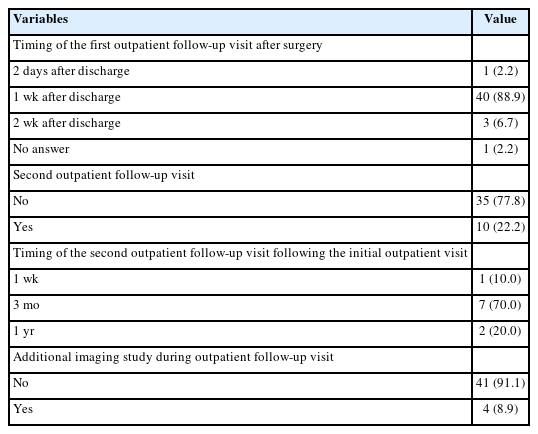
The Korean Association of Pediatric Surgeons (KAPS) performed a nationwide survey on sacrococcygeal teratoma in 2018.

Methods

The authors reviewed and analyzed the clinical data of patients who had been treated for sacrococcygeal teratoma by KAPS members from 2008 to 2017.

Results

A total of 189 patients from 18 institutes were registered for the study, which was the first national survey of this disease dealing with a large number of patients in Korea. The results were discussed at the 34th annual meeting of KAPS, which was held in Jeonju on June 21–22, 2018.

Conclusions

We believe that this study could be utilized as a guideline for the treatment of sacrococcygeal teratoma to diminish pediatric surgeons' difficulties in treating this disease and thus lead to better outcomes.

  • 10 View
  • 0 Download
[English]
Comparison of the Surgical Outcomes between Open Fundoplication and Laparoscopic Fundoplication in Children with Gastroesophageal Reflux Disease
Su young Hong, Hee-Beom Yang, Sao Thi Han, Hyun-Young Kim, Sung Eun Jung
Adv Pediatr Surg 2019;25(2):51-59.   Published online December 17, 2019
DOI: https://doi.org/10.13029/aps.2019.25.2.51
Purpose

This study aimed to compare the outcomes of open fundoplication (OF) and laparoscopic fundoplication (LF) in children with gastroesophageal reflux disease (GERD).

Methods

We retrospectively reviewed the electronic medical charts of pediatric patients who underwent fundoplication for GERD between January 2005 and May 2018 at the Korean tertiary hospital. Patient characteristics, operation type, associated diagnosis, operation history, neurologic impairment, postoperative complication, recurrence, and operation outcomes were investigated. The Mann-Whitney U test or Student's t-test was used to evaluate continuous data as appropriate. The χ2 test was used to analyze categorical data.

Results

A total of 92 patients were included in this study; 50 were male and 42 were female. Forty-eight patients underwent OF and 44 patients underwent LF. Patient characteristics, such as sex ratio, gestational age, symptoms, neurological impairment, and history of the previous operation were not different between the two groups. A longer operative time (113.0±56.0 vs. 135.1±49.1 minutes, p=0.048) was noted for LF. There was no significant difference in operation time when the diagnosis was limited to only GERD, excluding patients with other combined diseases. Other surgical outcomes, such as intraoperative blood loss, transfusion rate, hospital stay, and recurrence rate were not significantly different between the 2 groups. The complication rate was slightly higher in the OF group than in the LF group; however, the difference was not significant (20.8% vs. 11.4%, p=0.344).

Conclusion

LF is as safe, feasible, and effective as OF for the surgical treatment of GERD in children.

  • 6 View
  • 0 Download

Case Report

[English]
Recurrent Congenital Milium of the Nipple
Hyun Beak Shin, Yeon-Jun Jeong
Adv Pediatr Surg 2019;25(2):60-62.   Published online December 12, 2019
DOI: https://doi.org/10.13029/aps.2019.25.2.60

Congenital milia of the nipple are extremely rare, and standard treatment has not yet been established because nipple preserving excision is problematic due to the location. Although most reports show excisional biopsy resulting in good outcomes without recurrence, there is a lack of consensus about treatment modality, with several studies suggesting that incisional evacuation by needle, or a ‘wait and see’ approach represent sufficient treatment. This case report is about a recurrent case after incisional evacuation for congenital milium of the nipple. We recommend nipple preserving excision with exfoliation of the milial capsule as being the most appropriate treatment modality for congenital milium of the nipple.

  • 10 View
  • 0 Download

Original Article

[English]
Early Experience of Doxycycline Sclerotherapy for Lymphatic Malformations
Ju Yeon Lee, Jung-Man Namgoong, Seong Chul Kim, Dae Yeon Kim
Adv Pediatr Surg 2019;25(2):44-50.   Published online October 11, 2019
DOI: https://doi.org/10.13029/aps.2019.25.2.44
Purpose

Lymphatic malformations (LMs) are congenital malformations of the lymphatic system which can be effectively treated by sclerotherapy. This study aims to evaluate the efficacy of doxycycline in the treatment of LMs.

Methods

We retrospectively reviewed the medical records of all patients who were diagnosed as LMs and underwent doxycycline sclerotherapy in Asan Medical Center between March 2013 and February 2014. Thirty-five sclerotherapy procedures were performed on 21 patients. The procedures were performed under general anesthesia. After each treatment, the clinical and radiographic response was characterized as complete (≥80% decrease in lesion size), partial (<80% decrease of size), or no response (no decrease of size).

Results

There were 11 male patients and 10 female patients. The median age of sclerotherapy was 21 months (range, 2–180 months). The most common location was cervicofacial (52.3%), followed by extremity (28.6%) and truncal (19.0%). The most common lesion type was macrocystic (71.4%), followed by microcystic (28.5%). There was one (2.8%) skin necrosis which was recovered by wound management. Thirty-eight percent of patients had a complete response, 47.6% of patients had a partial response and 14.3% of patients had no response. Median frequency of treatment was one (range, 1–5). No response group consisted of all microcystic type.

Conclusion

Sclerotherapy with Doxycycline is safe and effective for macrocystic LMs.

  • 7 View
  • 0 Download

Case Report

[English]
A Case of Hemolytic Uremic Syndrome after Appendectomy
Layun Kim, Hye Lim Jung, Jung Yeon Shim, Deok Soo Kim, Jae Won Shim, Young Joo Han, Ji Hee Kwak
Adv Pediatr Surg 2019;25(1):29-34.   Published online July 3, 2019
DOI: https://doi.org/10.13029/aps.2019.25.1.29

Hemolytic uremic syndrome (HUS) is associated with consumption of under cooked gound beef, characterized by triad of renal failure, microangiopathic hemolytic anemia and thrombocytopenia. Early recognition of this disease, maintenance of fluid balance and proper dialysis seems to prevent acute mortality. A 23-month-old boy was admitted with abdominal pain, bloody diarrhea and fever. On hospital day (HD) #2, he developed aggravated abdominal pain compared to the initial assessment. Contrast abdominal computed tomography demonstrated findings suggestive acute appendicitis so the patient was underwent laparoscopic appendectomy. On HD #3 and #4, his laboratory findings showed marked thrombocytopenia and serum creatinine elevation. He was transferred to another hospital for dialysis with the impression of acute renal failure. Later, verotoxin-producing Escherichia coli, which is one of the most common causes of HUS, was detected on his initial stool examination. HUS can present with prodromal gastrointestinal symptoms mimicking acute abdomen. So even if the patient is primarily diagnosed acute abdomen in the initial setting, clinicians should always keep in mind of other fatal conditions disease, such as HUS. To our knowledge, this is the first case report of acute appendicitis followed by HUS in South Korea.

  • 5 View
  • 0 Download

Original Articles

[English]
A Comparative Study of Single Scrotal Incision Orchiopexy of Children with Palpable Low-Lying Undescended Testis with Traditional Inguinal Method
Davud Badbarin, Seyed Ehsan Mousavi Toomatari, Saeed Aslanabadi, Ebrahim Farhadi, Sara Akhavan Salamat
Adv Pediatr Surg 2019;25(1):14-19.   Published online July 1, 2019
DOI: https://doi.org/10.13029/aps.2019.25.1.14
Purpose

Undescended testis (UDT) is a developmental defect in which one or both testicles do not arrive in the scrotum. Its prevalence at birth and one year after is 2%–4% and less than 1%, respectively. Currently, surgery is recommended to treat congenital cryptorchidism in order to prevent testicular degeneration. Classic method is performed via incision in inguinal and scrotum and the new method is done via incision in scrotum.

Method

Sixty male participants with 65 UDT undergoing surgery were randomly assigned to scrotal incision (n=31) and classic inguinal incision methods (n=34). Patients were followed for 6 months and testicular atrophy, infection, recurrence, and duration of surgery were compared between two groups.

Results

Scrotal incision compared to classic incision method had significantly lower duration of surgery (19.06±2.96 minutes vs. 30±10.42 minutes; p=0.002) and recurrence during follow-up (0 vs. 5 cases; p=0.026). There was only one surgical site infection in the scrotal incision method. There were hematoma and post-operative swelling in 13.3% of cases after scrotal incision method.

Conclusion

Scrotal incision is an alternative method for the UDT with lower duration of surgery, lower recurrence rate, and better cosmetic results.

  • 12 View
  • 1 Download
[English]
Nonoperative Treatment of Appendicitis in the Pediatric Population: Stringent Patient Selection for Successful Application
Joonhyuk Son, Sanghoon Lee, Jiyoon Hong, Jeong-Meen Seo, Suk-Koo Lee
Adv Pediatr Surg 2019;25(1):7-13.   Published online June 28, 2019
DOI: https://doi.org/10.13029/aps.2019.25.1.7
Purpose

Although nonoperative treatment of appendicitis (NOTA) in the pediatric population has been well reported recently, patient selection and treatment scheme varies among studies, making it difficult to establish treatment standards for NOTA.

Methods

In a single medical center, patients younger than 18 years who were diagnosed with appendicitis: 1) with abdominal pain not exceeding 24 hours, 2) without radiologic evidence of appendicolith or appendiceal perforation or pelvic abscess, and 3) without signs of frank generalized peritonitis were offered NOTA, and their data were prospectively collected.

Results

Twenty-two patients with uncomplicated appendicitis agreed to NOTA and were enrolled in the study. The initial success rate (resolution of abdominal pain and hospital discharge without appendectomy) was 100% (22 out of 22 patients). At a median follow-up period of 23.8 months, two patients had recurrence at two and three months after completion of NOTA. These patients underwent laparoscopic appendectomy.

Conclusion

Stringent patient selection may be necessary to apply NOTA safely for all children with uncomplicated appendicitis. Further studies concerning patient selection and conformed treatment protocols for NOTA are required.

  • 8 View
  • 0 Download

Case Reports

[English]
Successful Correction of Long Gap Esophageal Atresia with Gastric Tube through Totally Laparoscopic and Thoracoscopic Procedure
Hee-Beom Yang, Hyun-Young Kim, Sung Eun Jung
Adv Pediatr Surg 2019;25(1):24-28.   Published online June 28, 2019
DOI: https://doi.org/10.13029/aps.2019.25.1.24

Esophageal atresia (EA) is a diverse disease entity. We present a case of long gap EA without fistula corrected through totally laparoscopic and thoracoscopic esophageal replacement using gastric tube. A male baby weighing 3,000 g, with suspicion of EA, was born at gestational age of 37+6 weeks. Gastrostomy was made at an age of two days; seven months later, definite operation was planned. We determined to perform the gastric tube replacement due to long gap revealed by fluoroscopy. Gastric mobilization, gastric tube formation, and pyloroplasty were performed laparoscopically. An isoperistaltic 9 cm gastric tube was made using 2 Endo GIA 45, and interrupted end-to-end esophago-esophagostomy was performed thoracoscopically. With laparoscopy, gastropexy to the diaphragm was performed through the interrupted suture. Operation time was 370 minutes; there was no intraoperative event. Postoperative course was uneventful. He underwent esophageal balloon dilatation due to anastomosis stenosis in the months after surgery.

  • 5 View
  • 0 Download
[English]
Congenital Anomaly of Ribs Masquerading as Diaphragmatic Hernia in an Infant: A Case Report
Priyanka Mittal, Shailesh Solanki, Prema Menon
Adv Pediatr Surg 2019;25(1):20-23.   Published online June 26, 2019
DOI: https://doi.org/10.13029/aps.2019.25.1.20

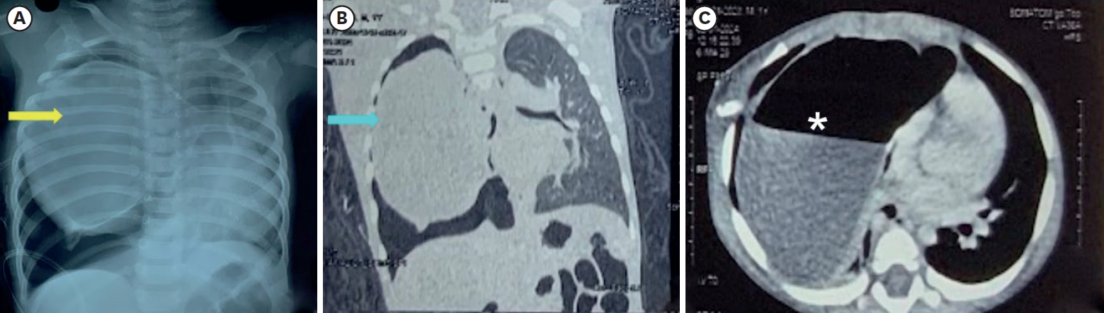
Congenital absence/hypoplasia/fusion of ribs are very rare anomalies and presentation varies from asymptomatic to life-threatening. Visibly evident cases are straightforward to diagnose but this is not always the case. Familiarity and awareness of these anomalies can help to diagnose cases with subtle signs and symptoms. Proper radiological investigations are vital for anatomical delineation. Absence or hypoplasia of the inferior ribs along with its attached muscles can cause ‘lung hernia’ that produces an unstable chest leading to paradoxical respiratory movements. Here, we present a case of hypoplastic and fused ribs in an infant, who presented with respiratory distress and created a diagnostic dilemma.

  • 13 View
  • 0 Download

Original Article

[English]
Purpose

The introduction of Malone antegrade continence enema) in the management of children with fecal incontinence has brought remarkable improvement in patient care, Malone originally described appendix as a conduit and it has become widely accepted. However, surgeons are faced with situations where appendix is not available, the selection and creation of other conduit is always a challenge. We present our technique and experience with the use of alternative catheterizable conduits for antegrade continence enema (ACE).

Methods

Retrospective review of children who underwent ACE procedure in our institution from March 2009 to January 2014. The details retrieved: indication, reason for non availability of appendix, type of conduit, complications and patient's satisfaction.

Results

Five children were identified in whom the appendix was not available or suitable. In four children cecal/colon-based flap was used and in one child, ileal (Monti) segment was used to create a conduit. The mean follow-up was 3.2 years. All patients were satisfied with the procedure and no stenosis or loss of conduit was noted in the follow-up.

Conclusion

Continent catheterizable conduit for ACE can be accomplished with transverse tubularized intestinal segments and cecal/colonic flaps, with excellent outcome, irrespective of tissue used. Surgeon's preference and the patient's peculiar anatomy should determine the surgical technique to be used.

  • 25 View
  • 1 Download

Review Article

[English]
Enterostomy Closure after Acute Abdomen in Neonate and Infant
Hee-Beom Yang, Hyun-Young Kim
Adv Pediatr Surg 2018;24(2):35-43.   Published online December 24, 2018
DOI: https://doi.org/10.13029/aps.2018.24.2.35

The risk of acute abdomen in neonates and infants is generally increased due to advanced maternal age and neonatal intensive care unit care development. Enterostomy is a safe procedure for acute abdomen in neonates and infants. However, there is no consensus for the optimal timing of enterostomy closure (EC). A few considerations should be reviewed for deciding the timing of stoma closure to obtain the best outcome. Distal loopography is commonly performed upon examination to ascertain the existence of a distal passage after EC, detect signs of disease-specific complications, and assess the need of surgery in addition to EC. Pathology review is also one important pre-closure consideration. When the incidence of a hypoganglionosis or an aganglionosis combined with acute abdomen is observed in neonates and infants, thorough examinations should be performed to conclusively determine whether there is no passage disturbance of the distal bowel. Refeeding not only provides information about chance to grow, electrolyte imbalance correction, and proximal and distal bowel size match, but also provides information about distal bowel passage when there is a doubtful distal loopography or pathology result. Early closure enables growth spurt with the correction of water/electrolyte imbalance. It potentially reduced medical costs, less discrepancy between proximal and distal bowel size. Some factors favoring a late stoma closure may be due to less postoperative complications, early recovery, shorter time of total parenteral nutrition after EC, and decreased length of hospitalization after the EC. Some studies have shown controversial results. In summary, a pre-closure evaluation is imperative to assure the safety of an EC. The optimal timing of an EC remains controversial. For this reason, individualized approach is needed after reviewing the general condition of each patient. Further prospective study on optimal timing of stoma closure including a randomized clinical trial is needed.

  • 23 View
  • 0 Download

Original Article

[English]
Abdominal Lymphatic Malformation in Children
Sangho Lee, Jinyoung Park
Adv Pediatr Surg 2018;24(2):60-67.   Published online December 21, 2018
DOI: https://doi.org/10.13029/aps.2018.24.2.60
Purpose

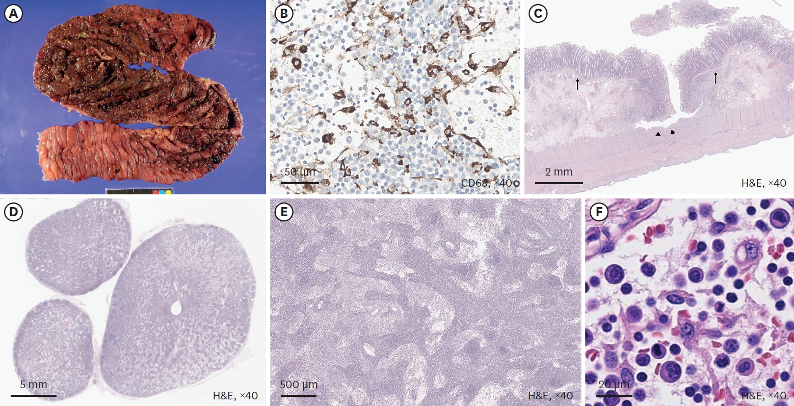
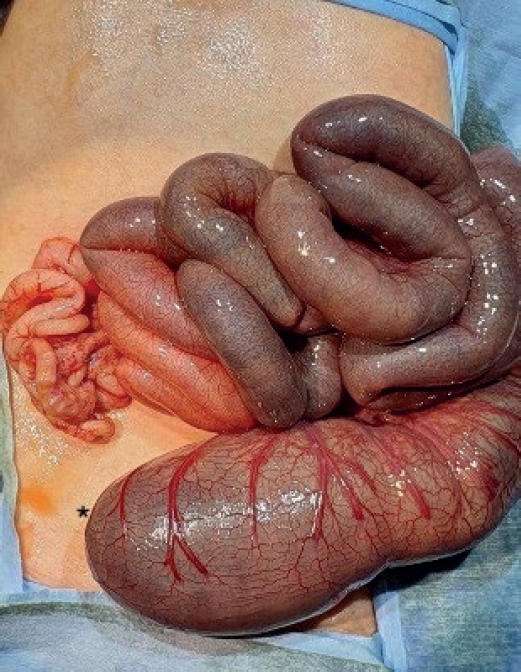
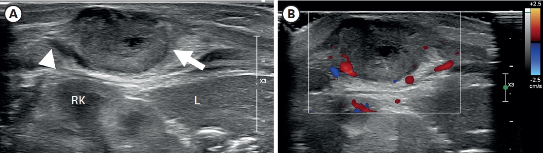
Lymphatic malformations are benign congenital malformations of the lymphatic system that occur predominantly in children. Most lymphatic malformations occur in the head and neck region, with those in abdominal locations, such as the mesentery, omentum and retroperitoneum, being less common, accounting for fewer than 5% of lymphatic malformations in children. This study analyzed the clinicopathologic characteristics and treatment outcomes of abdominal lymphatic malformations in children.

Methods

The medical records of 12 pediatric patients treated for abdominal lymphatic malformations at our institution between April 1999 and September 2017 were retrospectively reviewed. Demographic and clinical characteristics, including gender, age, symptoms and signs, diagnostic modalities, and treatment results, were analyzed.

Results

The 12 patients included 11 boys and 1 girl, ranging in age from 3 months to 17 years (median 36.5 months) at presentation. The primary signs and symptoms included abdominal pain, abdominal mass and abdominal distention. Other symptoms and signs included fever, vomiting, scrotal pain and mass, and right inguinal mass. All patients were diagnosed by abdominal ultrasonography and computed tomography, and all underwent surgical excision with or without bowel resection. The lymphatic malformations occurred in the retroperitoneum (n=4), omentum (n=4), jejunal mesentery (n=2), and retroperitoneum and mesentery (n=2). Seven patients underwent complete mass excision, including 3 who underwent laparoscopic excisions, and 3 who underwent mass excision with segmental resection of the adjoining bowel. Two patients underwent incomplete excision because the lesion was extensive and invaded the superior mesenteric vessels. There was no major perioperative morbidity in any patient. At a median follow-up of 50 months (range, 8–183 months), only 1 patient experienced recurrence.

Conclusion

Although abdominal lymphatic malformations are benign, most children present with acute abdominal symptoms, necessitating early surgical treatment.

  • 5 View
  • 0 Download

Case Report

[English]
Two Cases of Pyriform Sinus Cyst in Neonates: Importance of Early Recognition and Differential Diagnosis
Mi Hae Chung, So Young Kim, Gye-Yeon Lim, Jae Hee Chung
Adv Pediatr Surg 2018;24(2):100-106.   Published online December 13, 2018
DOI: https://doi.org/10.13029/aps.2018.24.2.100

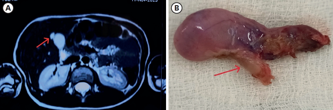
Early suspicion is essential in diagnosing pyriform sinus cysts. We report two neonatal cases of pyriform sinus cysts presented as neck masses. The first case presented as a right neck mass, which made it more difficult to suspect a pyriform sinus cyst considering the prevalence of left sided cysts. Surgical resection was done in both cases and anatomical investigation suggested both to originate from the fourth branchial pouch. Detection of air bubble containing mass on imaging studies can aid early diagnosis and early use of gastric tube feeding can facilitate treatment by preventing milk contamination which may result in infection of the sinus cyst.

  • 11 View
  • 0 Download

Original Article

[English]
Perioperative Outcomes and Surgical Indications of Minimally Invasive Pancreatectomy for Solid Pseudopapillary Tumor in Pediatric Patients
Chong Won Lee, Jung-Man Namgoong, Dae Yeon Kim, Seong Chul Kim, Soo Young Lee, Yujeong Cho, Hyunhee Kwon
Adv Pediatr Surg 2018;24(2):76-85.   Published online December 10, 2018
DOI: https://doi.org/10.13029/aps.2018.24.2.76
Purpose

We evaluated perioperative and long-term outcomes of minimally invasive surgery (MIS) and established indications of MIS in solid pseudopapillary tumor (SPT) in pediatric patients.

Methods

From October 1992 to April 2018, 66 patients (age, <18 years) diagnosed with SPT underwent either open pancreatectomy (OP) or MIS. Variables including postoperative complications and recurrence rates were retrospectively analyzed.

Results

Thirty-five patients underwent open surgery and 31 underwent laparoscopic/robotic surgery. Mean tumor size in MIS was significantly smaller than that in OP (4.3±1.8 cm vs. 7.6±3.5 cm, p=0.005). There were 4 cases of open conversion from laparoscopic surgery because of vessel encasements (n=2), bleeding (n=1), and pancreatic ductal injury (n=1). Solitary pseudopapillary carcinoma was diagnosed in 6 patients. Recurrence was observed in 3 and 1 patients who underwent OP and MIS, respectively (p=0.634). Tumor size, mass size/abdominal diameter (MS/AD) ratio, and degree of the portal or superior mesenteric vein involvement were the most important indications for MIS.

Conclusion

MIS is being widely used in pediatric surgeries with increased expertise and safety, especially in pancreatic diseases. Careful patient selection for MIS in regards with parameters such as MS/AD ratio and vessel abutment might be a feasible choice.

  • 5 View
  • 0 Download

Case Report

[English]
Prolonged Gallbladder Hydrops in a Kawasaki Disease Patient
Kyung Lim Yoon, Do Hee Kim, Mi Young Han, Sung Ho Cha, Hyun Cheol Kim
Adv Pediatr Surg 2018;24(2):107-112.   Published online December 10, 2018
DOI: https://doi.org/10.13029/aps.2018.24.2.107

Although gallbladder (GB) hydrops in childhood is uncommon, it is most commonly associated with Kawasaki disease (KD) and can be a risk factor of poor outcomes in patients with KD. Our patient presented with a fever, diffuse abdominal pain, maculopapular rash, bilateral conjunctivitis, red and fissured lip tissue, and 3 days of right cervical swelling. Abdominal examination revealed abdominal distension and tenderness. Abdominal ultrasonography was performed to identify the source of abdominal symptoms, which revealed dilatation of the GB. Abdominal computed tomography scan was also performed to exclude other hepatobiliary disease, which showed normal hepatobiliary systems. The GB of our patients returned to a normal size without any complications, but it regressed very slowly. We report a case of a 6-year-old boy with KD who presented with markedly distended and unusually prolonged GB hydrops.

  • 7 View
  • 0 Download

Original Articles

[English]
Effect of Nutritional Support on Postoperative Growth Velocity of Neonates in Neonatal Intensive Care Unit
Hee-In Jo, Ji-Young Sul, Jun-Beom Park
Adv Pediatr Surg 2018;24(2):51-59.   Published online December 9, 2018
DOI: https://doi.org/10.13029/aps.2018.24.2.51
Purpose

This study aimed to evaluate the relationship between nutritional support and growth velocity after abdominal surgery in neonates.

Methods

The electronic medical records of 45 neonates who underwent abdominal surgery in neonatal intensive care unit from 2012 to 2016 were collected to see how surgery and postoperative nutrition affect for the growth of neonate with abdominal surgery. The growth velocity was measured from the time of surgery to the time of discharge based on body weight.

Results

In neonates who achieve their protein requirement on the first day after surgery, the growth velocity was better than that in neonates who did not achieve their protein requirement on the first day after surgery (4.31 vs. 15.21; p=0.004). Based on the type of surgery, length of bowel resection and surgical complications, this study showed better growth velocity in neonates who had no surgical complications (5.34 vs. 12.74; p=0.775), reoperation (5.25 vs. 22.19, p=0.987), or bowel resection (6.79 vs. 9.95, p=0.302). However, there was no statistically significant difference among these factors.

Conclusion

We concluded in this study that adequate protein supplement from the first day of surgery could have a positive effect on the growth velocity of neonates who underwent abdominal surgery.

  • 7 View
  • 0 Download
[English]
Pneumothorax Is a Significant Risk Factor for Mortality in Congenital Diaphragmatic Hernia: A Single Center Experience
SeungWook Han, Sanghoon Lee, Joonhyuk Son, Jeong-Meen Seo, Suk-Koo Lee
Adv Pediatr Surg 2018;24(2):68-75.   Published online December 7, 2018
DOI: https://doi.org/10.13029/aps.2018.24.2.68
Purpose

We describe our experience in managing congenital diaphragmatic hernia (CDH) in neonates.

Methods

From February 1995 to July 2014, 64 neonates diagnosed with CDH were managed. The medical records of these neonates were retrospectively reviewed.

Results

There were 40 males and 24 females. CDH was on the left side in 44 cases (68.8%), on the right side in 19 cases (29.7%) and bilateral in 1 case (1.6%). Forty-six patients (71.9%) received surgical repair of the hernia while 18 patients (28.1%) died prior to surgery. The timing of surgery was mean 7.0 days after birth. Extracorporeal membrane oxygenation was used in six patients (9.4%), High-frequency oscillation ventilation was used in 49 patients (76.6%), and nitric oxide was used in 42 patients (65.6%). Thirty-three cases (71.7%) of CDH repair were done via open laparotomy, 3 cases (6.6%) by open thoracotomy. and minimally invasive thoracoscopic repair was done in 10 cases (21.7%). Ten cases (21.8%) required patch repair of the CDH. Barotrauma and pneumothorax of the contralateral lung was seen in 16 cases, leading to death in 15 of these cases. The overall survival rate was 48.4% (31/64) and postoperative survival rate was 67.4% (31/46). When all patients are divided into 3 groups by era and analyzed by logistic regression models, the mortality outcome of recent era (2009–2014) was significantly better than that of intermediate era (2002–2008) (29% vs. 71%, p=0.006) and tended to be better than that of past era (1995–2001) (42% vs. 71%, p=0.062).

Conclusion

The overall survival of neonates with CDH at our center has improved over the last two decades. Sixty-four neonates with CDH were managed at a single center and their overall survival was 48.4%. The risk factors for mortality include the occurrence of pneumothorax and right side lesions.

  • 9 View
  • 0 Download

Case Report

[English]
Double H-type Tracheoesophageal Fistulae: A Case Report
Joohyun Sim, Jeong Hong
Adv Pediatr Surg 2018;24(2):94-99.   Published online December 3, 2018
DOI: https://doi.org/10.13029/aps.2018.24.2.94

We present an extremely rare case with double H-type tracheoesophageal fistulae identified with a time-lapse and repaired separately. A newborn male presented with cyanosis after breastfeeding. Contrast esophagogram demonstrated an H-type fistula, and then it was repaired in a standard fashion via right thoracotomy. When routine esophagogram was taken on postoperative day 10, another fistula was noticed at a level higher than the previous one. Bronchoscopy was performed to evaluate the lesion whether it was a recurred fistula or a second H-type fistula. However, it was so tiny that it was not visible with bronchoscopy. It was discovered only two months later when the fistula had grown up with the baby. The second H-type fistula was repaired through a cervical incision. Although double H-type fistulae are extremely rare, the possibility of another fistula, as well as recurrence, must be ruled out when symptom recurs after a definitive operation of an H-type fistula.

  • 10 View
  • 0 Download

Original Article

[English]
Efficacy and Safety of Single-Site Umbilical Laparoscopic Surgery for Small Bowel Resection in Pediatric Patients
Kang-Ho Lee, Soo-Hong Kim, Yong-Hoon Cho, Hae-Young Kim
Adv Pediatr Surg 2018;24(2):44-50.   Published online December 3, 2018
DOI: https://doi.org/10.13029/aps.2018.24.2.44
Purpose

Single-port laparoscopy-assisted surgery is being performed for various operations in pediatric patients recently. The aims of this study were to prove the safety and find well-matched indications of small bowel resection using single-site umbilical laparoscopic surgery (SSULS).

Methods

From 2011 to 2016, 29 pediatric patients underwent SSULS. Medical records were retrospectively reviewed.

Results

A total of 29 patients were included and 30 SSULS were performed in this study. The mean age at operation was 5.7 years, and the mean weight was 21.9 kg. Meckel's diverticulum was the most common indication for SSULS, followed by small bowel intussusception due to leading point mass, small bowel duplication, and Crohn's disease. In most cases, estimated blood loss was negligible except in Crohn's disease with severe inflammation. While answering post-discharge questions about scars, most parents responded that they were satisfied with the postoperative wound.

Conclusion

SSULS is a useful operation to try even for surgeons who do not have advanced laparoscopic skills. Complication rates of single-port operations do not differ from those of conventional laparoscopic operations. Most lesions of the small bowel could be indications of SSULS. Careful attention is required when performing SSULS in patients with Crohn's disease.

  • 19 View
  • 0 Download

Case Report

[English]
Long-Term Outcome of Patients Undergoing Total Proctocolectomy with Ileal Pouch-Anal Anastomosis in Childhood
Yoon-Jung Sim, Chaeyoun Oh, Joong Kee Youn, Soo-Hong Kim, Kyu Joo Park, Seung-Yong Jeong, Sung-Eun Jung, Kwi-Won Park, Hyun-Young Kim
Adv Pediatr Surg 2018;24(2):86-93.   Published online November 26, 2018
DOI: https://doi.org/10.13029/aps.2018.24.2.86
Purpose

Total proctocolectomy with ileal pouch-anal anastomosis (T-IPAA) in childhood is a surgical procedure mainly applied to familial adenomatous polyposis (FAP) or ulcerative colitis (UC), but it can be applied to non-FAP/non-UC disease (NFNU). Studies regarding the role of T-IPAA who underwent the operation in childhood, especially in terms of long-term gastrointestinal function, complications, and quality of life (QOL) are limited. The aim of this study was to evaluate the characteristics of patients receiving T-IPAA and to compare their bowel function outcomes and QOL.

Methods

Patients aged ≤18 years at the time of T-IPAA were included. Their medical records were retrospectively reviewed. Krickenbeck classification, Cleveland Clinic Incontinence (CCI) score, 36-item Short-form Health Survey Questionnaire, and Gastrointestinal Quality of Life Index were used for the evaluation of bowel function and QOL. The median follow-up period was 9.8 years.

Results

Of the 25 patients, 9 had FAP, 9 had UC, and 7 had NFNU. NFNU include 3 of Hirschsprung disease, 2 of intestinal neuronal dysplasia, and 2 of imperforate anus. The median age at T-IPAA was 17.8, 14.2, and 9.3 years for FAP, UC, and NFNU, respectively (p=0.001). Bowel function was satisfactory in terms of voluntary bowel movement (VBM), soiling, and constipation. VBM and constipation were not different between the groups, but soiling was most in NFNU (100%, p=0.047). However, QOL was best in the NFNU group in surveys (p=0.034 and 0.004, respectively).

Conclusion

T-IPAA could be safely applied not only for FAP and UC but also for other diseases in selective cases, with caution.

  • 5 View
  • 0 Download

Original Article

[English]
Is Preoperative Ultrasonography Necessary in Pediatric Inguinal Hernia?
Ji-Won Han, Joong Kee Youn, Hee-Beom Yang, Chaeyoun Oh, Hyun-Young Kim, Sung-Eun Jung
J Korean Assoc Pediatr Surg 2018;24(1):5-9.   Published online June 30, 2018
DOI: https://doi.org/10.13029/jkaps.2018.24.1.5
Purpose

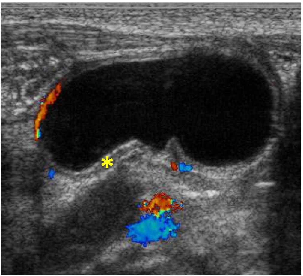
Preoperative ultrasonography (USG) in pediatric inguinal hernia has controversy. In this study, we analyzed the cases of pediatric inguinal hernia with/without preoperative USG and discussed whether USG is necessary.

Methods

We reviewed medical records of 1,441 patients who underwent inguinal hernia repair in Seoul National University Children's Hospital between January 2011 and August 2016 retrospectively.

Results

Male were 69.3% and age at operation was 37.8±36.5 months old. There were 150 patients (10.4%) performed USG preoperatively. The department ordered to perform USG included department of surgery (n=71), emergency medicine (n=42), pediatrics (n=26), urology (n=10) and outside hospital (n=1). The reasons of performing USG included evaluation for hernia laterality (n=82), incarceration (n=28), testis (n=15), request of parents (n=14), scrotal mass (n=6) and incidentally found during evaluation for another disease (n=5). Excepting 5 cases of incidental finding, of 145 cases with USG, 12 (8.3%) cases changed the surgical plan; change to bilateral repair from unilateral repair (n=5), emergency operation due to incarceration (n=4) which include 1 salpingo-oophorectomy, 1 open abdomen surgery and 2 hernia repair after reduction of ovary, change to co-operation of orchiopexy (n=2) and change to laparoscopic surgery from open surgery due to herniation of both ovaries into one inguinal canal (n=1). In group without USG (n=1,291), 5 patients (0.4%) had unexpected problems during operation; 2 co-operation of orchiopexy because of transverse testicular ectopia (n=1) and right undescended testis (n=1), 2 hypertrophy of major labia and 1 retroperitoneal lymphangioma at inguinal area misdiagnosed as inguinal hernia.

Conclusion

It was difficult to interpret the meaning of preoperative USG because not all patients had performed it. In this study, 10.4% of patients performed USG and 8.3% of them changed surgical plan. About 0.4% of patients without preoperative USG would have benefited from it for surgery if they had performed it. Since the percentage is too low, it is unreasonable to conclude that USG has diagnostic utility in inguinal hernia in this study.

  • 4 View
  • 0 Download

Case Reports

[English]
Bile Duct Stricture and Intrahepatic Cystic Formation after Abdominal Injury due to Child Abuse: A Case Report
Kyong Ihn, Eun-Jung Koo, In Geol Ho, Dongeun Lee, Seok Joo Han
J Korean Assoc Pediatr Surg 2018;24(1):30-34.   Published online June 30, 2018
DOI: https://doi.org/10.13029/jkaps.2018.24.1.30

A 6-year-old male who lived with a mother in a single-parent family was referred to the emergency room with multiple traumas. There was no specific finding on CT scan of the other hospital performed 55 days before admission. However, CT scan at the time of admission showed common bile duct (CBD) stenosis, proximal biliary dilatation and bile lake formation at the segment II and III. Endoscopic retrograde biliary drainage was performed, but the tube had slipped off spontaneously 36 days later, and follow-up CT scan showed aggravated proximal biliary dilatation above the stricture site. He underwent excision of the CBD including the stricture site, and the bile duct was reconstructed with Roux-en-Y hepaticojejunostomy. Pathologic report of the resected specimen revealed that the evidence of trauma as a cause of bile duct stricture. While non-iatrogenic extrahepatic biliary trauma is uncommon, a level of suspicion is necessary to identify injuries to the extrahepatic bile duct. The role of the physicians who treat the abused children should encompass being suspicious for potential abdominal injury as well as identifying visible injuries.

  • 7 View
  • 0 Download
[English]
Transverse Colon Volvulus around the Gastrostomy Tube in a Pediatric Situs Inversus Patient
Yoon Hyung Kang, Joong Kee Youn, Ji-Won Han, Chaeyoun Oh, Sung-Eun Jung, Hyun-Young Kim
J Korean Assoc Pediatr Surg 2018;24(1):26-29.   Published online June 30, 2018
DOI: https://doi.org/10.13029/jkaps.2018.24.1.26

An 18-year-old male patient with cerebral palsy and scoliokyphosis came to the emergency department with abdominal distension and vomiting. He was a situs inversus patient with a feeding gastrostomy tube. Sigmoid volvulus was initially suspected, so rectal tube insertion and endoscopic decompression were attempted, but failed. So he went through explorative laparotomy, and transverse colonic adhesion and twisting around the gastrostomy tube and gastric wall was identified. Adhesiolysis and resection with redundant transverse colon and end-to-end colocolic anastomosis was performed. He discharged with symptom free. Suspecting transverse colonic volvulus is important when the patient has anatomical anomalies and feeding gastrostomy tube. Timely diagnosis with proper radiologic imaging should be made. Surgical resection of the redundant colon is needed for successful management of transverse colonic volvulus.

  • 6 View
  • 0 Download

Original Articles

[English]
Santulli Enterostomy: A Considerable Method for Patients Who Require Proximal Enterostomy
Kyong Ihn, Eun-Jung Koo, In Geol Ho, Seok Joo Han, Jung-Tak Oh
J Korean Assoc Pediatr Surg 2018;24(1):20-25.   Published online June 30, 2018
DOI: https://doi.org/10.13029/jkaps.2018.24.1.20
Purpose

Santulli enterostomy has been used for various surgical abdominal conditions that require temporary diversion of bowel during a neonatal period. The aim of this study was to report clinical outcomes of Santulli enterostomy and to evaluate its usefulness.

Methods

Between January 2000 and December 2016, 40 neonates who underwent Santulli enterostomy were enrolled; Santulli enterostomies were performed for 25 patients without previous laparotomy (primary Santulli group) and 15 patients with previous laparotomy (secondary Santulli group).

Results

Small bowel atresia is the first common indication of Santulli enterostomy (22/40, 55.0%), and luminal discrepancy between proximal and distal bowel was the most common determinant factor of Santulli enterostomy (17/40, 42.5%). The median age at surgery and mean birth weight were 2 days and 2,480 g respectively in the primary group, and 71 days, 2,340 g respectively in the secondary group. Operation time was significantly longer in the secondary group than the primary group (156±48 minutes vs. 224±95 minutes, p=0.019), and there was no difference in the time taken to initiation of oral feeding between the two groups. Santulli enterostomy closure was performed at median 65 days after Santulli enterostomy for primary group and 70 days for secondary group. Six complications (15.0%) were found after Santulli enterostomy, and nine complications (24.3%) after Santulli enterostomy closure (p=0.302). The incidence of complications was significantly higher in secondary group than in primary group (4.5% vs. 53.3%, p=0.001), and the reoperation rate was also significantly higher in the secondary group (4.5% vs. 46.7%, p=0.004).

Conclusion

Santulli enterostomy could be applied as a temporary enterostomy in neonatal patients with various surgical abdominal diseases. Considering the high complication rate after secondary Santulli enterostomy closure, decision making on the timing of enterostomy closure should be done with caution.

  • 7 View
  • 0 Download
[English]
The Treatment Experience of Lymphatic Malformations in Pediatric Patients
Jae Ha Park, So Hyun Nam
J Korean Assoc Pediatr Surg 2018;24(1):14-19.   Published online June 30, 2018
DOI: https://doi.org/10.13029/jkaps.2018.24.1.14
Purpose

The management of lymphatic malformation (LM) in pediatric patients is challenging. Complete excision of LM is difficult to achieve in some cases. We reviewed our experience how to manage LM.

Methods

We retrospectively reviewed the patients who were treated for LM between 2010 and 2017. Medical records were reviewed about age of diagnosis, age of treatment age, gender, symptom, location of tumor, treatment modality, response and complication.

Results

Sixty-three patients (39 boys and 24 girls) were included. Mean age at diagnosis was 14.5±28.0 months (range, neonate-10 years). The involved lesion were head and neck in 27 patients (42.9%), abdominal cavity in 7 patients (11.1%), chest wall and abdominal wall in 11 patients (17.5%), buttock in 7 patients (11.1%), and extremities in 11 patients (17.5%). The treatment options were including surgical resection in 32 patients, sclerotherapy in 7 patients, surgical resection combined sclerotherapy in 19 patients, and close observation in 5 patients. We achieved complete remission in 39 patients. Fourteen patients showed partial remission and 6 showed recurrences.

Conclusion

Despite surgical difficulty, meticulous excision with supportive treatment, and adjuvant sclerotherapy could get a favorable outcome without fatal complication. Decision should be based on surgeon's experience, location of LM, related symptoms, and consultation with patient's parents.

  • 5 View
  • 0 Download
[English]
Is Barium Enema Reliable for the Diagnosis of Total Colonic Aganglionosis?
Ju Yeon Lee, Dae Yeon Kim, Jeong Rye Kim, Jung-Man Namgoong, Seong Chul Kim
J Korean Assoc Pediatr Surg 2018;24(1):10-13.   Published online June 30, 2018
DOI: https://doi.org/10.13029/jkaps.2018.24.1.10
Purpose

Barium enema is one of the diagnostic modalities for Hirschsprung'sdisease. The present study aimed to investigate the diagnostic accuracy of barium enema for Hirschsprung's disease, especially total colonic aganglionosis (TCA).

Methods

We retrospectively reviewed the medical records of all the patients who were diagnosed as having TCA and underwent a barium enema in Asan Medical Center Children's Hospital between January 1998 and December 2016. All the tests were performed and reviewed by pediatric radiologists.

Results

Among the total 19 patients with TCA who underwent barium enema, 9 patients (47.4%) had accurate radiographic results. Eight of the 13 neonate patients (61.5%) showed typical TCA radiological findings. However, only one of the 6 patients aged >4 weeks (16.7%) had accurate radiological diagnosis.

Conclusion

Barium enema showed low accuracy for TCA, and its diagnostic performance was better in neonatal period than in those aged >4 weeks.

  • 7 View
  • 0 Download

Review Article

[English]
Congenital Esophageal Stenosis in Children: From Etiology to Prognosis
Soo-Hong Kim, Hyun-Young Kim
J Korean Assoc Pediatr Surg 2018;24(1):1-4.   Published online June 30, 2018
DOI: https://doi.org/10.13029/jkaps.2018.24.1.1

Congenital esophageal stenosis (CES) is a rare disease that has been reported to occur once in every 25,000 to 50,000 births. According to its etiology, CES is divided into 3 subtypes, tracheobronchial remnants (TBR), fibromuscular hypertrophy (FMH) and membranous diaphragm (MD). Symptoms begin at the weaning period and the introduction of solid food around 6 months with dysphagia and vomiting. Esophagography is first screening test and endoscopic ultrasonography plays important roles to diagnose subtypes deciding therapeutic plan. TBRs were generally treated with surgical resection and end-to-end anasotomosis, whereas FMH and MD had good response rate to endoscopic or radiologic guided dilatation. This article reviews the literature on the etiology, clinical course, diagnosis and management of CES including recent opinion.

  • 17 View
  • 0 Download