Skip to main navigation Skip to main content
  • KAPS
  • E-Submission

APS : Advances in Pediatric Surgery

OPEN ACCESS
ABOUT
BROWSE ARTICLES
EDITORIAL POLICY
FOR CONTRIBUTORS

Page Path

10
results for

"Soo Jin Na Choi"

Article category

Keywords

Publication year

Authors

Funded articles

"Soo Jin Na Choi"

Original Articles

[English]
Neuroblastoma: A 2020 Survey Conducted by the Korean Association of Pediatric Surgeons
Hee-Beom Yang, Soo Min Ahn, Min Jeng Cho, Yong-Hoon Cho, Soo Jin Na Choi, Yoon Mi Choi, Jae Hee Chung, Seok Joo Han, In Geol Ho, Jeong Hong, Kyong Ihn, Yeon Jun Jeong, Eunyoung Jung, Dae Youn Kim, Hae-Young Kim, Ki Hoon Kim, Seong Chul Kim, Soo-Hong Kim, Eun-Jung Koo, Hyun Hee Kwon, Yong Jae Kwon, Nam-Hyuk Lee, Ju Yeon Lee, Sanghoon Lee, Jung-Man Namgoong, Chaeyoun Oh, Jung-Tak Oh, Jin Young Park, Junbeom Park, Jeong-Meen Seo, Jae Ho Shin, Hyun Beak Shin, Joohyun Sim, Jiyoung Sul, Joon Kee Youn, Hyun-Young Kim
Adv Pediatr Surg 2025;31(2):66-76.   Published online November 25, 2025
DOI: https://doi.org/10.13029/aps.2025.31.2.66
Purpose
To report a nationwide survey on neuroblastoma conducted by the Korean Association of Pediatric Surgeons (KAPS) in 2020.
Methods
The clinical data of pediatric patients diagnosed with and treated for neuroblastoma from 2005 to 2019 in 19 hospitals of KAPS members were collected. Survival and prognostic factor analyses were performed using the log rank test and Cox proportional hazard analysis. A p-value <0.05 was considered significant.
Results
A total of 669 patients with neuroblastoma were registered for the study. The results were presented and discussed at the 36th annual meeting of the KAPS, which was held in Seoul on August 21, 2020.
Conclusion
This study provides information on patient demographics, prognostic outcomes, and comprehensive treatment outcomes for neuroblastoma. The study is expected to be an important reference for improving pediatric surgeons’ understanding and treatment of neuroblastoma.
  • 26 View
  • 2 Download
[English]
A Comparative Study of Three National Surveys on Biliary Atresia by the Korean Association of Pediatric Surgeons
Yeon Jun Jeong, Dayoung Ko, Hyunhee Kwon, Ki Hoon Kim, Dae Yeon Kim, Soo-Hong Kim, Wontae Kim, Hae-Young Kim, Hyun Young Kim, Seong Chul Kim, Younghyun Na, Jung-Man Namgoong, So Hyun Nam, Junbeom Park, Jinyoung Park, Tae-Jun Park, Jeong-Meen Seo, Ji-Young Sul, Joonhyuk Son, Hyun Beak Shin, Joohyun Sim, Soo Min Ahn, Hee Beom Yang, Jung-Tak Oh, Chaeyoun Oh, Joong Kee Youn, Sanghoon Lee, Ju Yeon Lee, Kyong Ihn, Hye Kyung Chang, Eunyoung Jung, Jae Hee Chung, Yu Jeong Cho, Yun Mee Choe, Soo Jin Na Choi, Seok Joo Han, In Geol Ho, Ji-Won Han
Adv Pediatr Surg 2025;31(2):47-58.   Published online July 16, 2025
DOI: https://doi.org/10.13029/aps.2025.31.2.47
Purpose
Biliary atresia (BA) is a rare but progressive cholangiopathy and the leading cause of pediatric liver transplantation worldwide. The Korean Association of Pediatric Surgeons (KAPS) has conducted three national surveys (2001, 2011, and 2023) to assess long-term trends in the diagnosis, treatment, and outcomes of BA. This study provides a comparative analysis of the 2nd and 3rd national surveys, with reference to selected findings from the 1st survey.
Methods
This study included 453 patients from the 3rd national survey (2011–2021) and 435 patients from the 2nd survey (2001–2010), all of whom underwent Kasai portoenterostomy. Data were collected via electronic case report forms from pediatric surgical centers nationwide. Comparisons were made regarding demographics, clinical features, diagnostic patterns, operative details, follow-up outcomes, and survival. Kaplan–Meier analysis was used to evaluate long-term survival.
Results
The mean number of BA patients per year remained stable between surveys (43.5 in the 2nd, 41.18 in the 3rd), though centralization of care increased, with 61.5% of cases managed by two major institutions in the 3rd survey. The median age at surgery decreased, and the use of preoperative imaging (especially magnetic resonance cholangiopancreatography) increased. The 10-year native liver survival rate declined from 59.8% to 53.7%, while overall 10-year survival improved slightly (92.9% to 93.2%). Postoperative complications, such as cholangitis and liver failure, persisted but were better categorized. The 3rd survey also reported improved mortality (4.9%) and reduced follow-up loss (11.5%) compared to the 2nd survey.
Conclusion
While overall survival after Kasai operation has remained high and even improved, native liver survival has slightly declined. The findings reflect earlier diagnosis, more consistent diagnostic imaging, and increasing centralization of care. These trends underscore the importance of long-term nationwide data collection in guiding future strategies for BA management in Korea.
  • 35 View
  • 4 Download
[English]
National Survey of Gastroschisis and Omphalocele by Korean Association of Pediatric Surgeons
Yeon Jun Jeong, Dayoung Ko, Eun-Jung Koo, Hyunhee Kwon, Dae Yeon Kim, Soo-Hong Kim, Wontae Kim, Hae-Young Kim, Hyun Young Kim, Seong Chul Kim, Younghyun Na, Jung-Man Namgoong, So Hyun Nam, Sungjoo Park, Junbeom Park, Jinyoung Park, Tae-Jun Park, Jeong-Meen Seo, Ji-Young Sul, Joonhyuk Son, Hyun Beak Shin, Joohyun Sim, Jung-Tak Oh, Chaeyoun Oh, Joong Kee Youn, Sanghoon Lee, Ju Yeon Lee, Cheolgu Lee, Kyong Ihn, Eunyoung Jung, Jae Hee Chung, Yong-Hoon Cho, Yun Mee Choe, Soo Jin Na Choi, Seok Joo Han, In Geol Ho
Adv Pediatr Surg 2024;30(2):39-51.   Published online December 13, 2024
DOI: https://doi.org/10.13029/aps.2024.30.2.39
Purpose
This study provides insights into the prevalence at birth, clinical characteristics, and outcomes of gastroschisis and omphalocele in Korea over the past decade, addressing the lack of localized data despite advanced healthcare capabilities.
Methods
The study retrospectively analyzed data from 20 pediatric surgical centers in Korea from January 2012 to December 2021, including 269 patients diagnosed with gastroschisis or omphalocele. Data variables included gender, gestational age, birth weight, associated anomalies, type of defect, surgical interventions, and outcomes.
Results
The study covered 269 patients, with 80 gastroschisis and 189 omphalocele cases. Gastroschisis prevalence at birth remained stable at 2.15 per 100,000 live births, while omphalocele increased to 5.08 per 100,000. Both conditions had similar gender ratios (0.95). Gastroschisis patients had lower birth weights (2,463.90±505.50 g) and smaller head circumferences (31.97±1.86 cm) compared to omphalocele patients (2,757.65±761.24 g, 32.78±2.64 cm). Omphalocele cases had more associated anomalies, especially cardiovascular issues. Prenatal diagnosis rates were high: 93.7% for gastroschisis and 86.4% for omphalocele. About 96.3% of gastroschisis and 84.1% of omphalocele patients were born in their treatment hospitals. Gastroschisis patients underwent surgery sooner (average 3.5 days) and started feeding later (16.5 days) than omphalocele patients (average 56.5 days to surgery, 6.6 days to start feeding). Hospital stays and follow-up durations were similar, averaging around 782.6 days for gastroschisis and 800.3 days for omphalocele patients. Survival rates were 89.7% for gastroschisis and 87.1% for omphalocele.
Conclusion
The study highlights the need for early diagnosis, centralized care, and specialized surgical approaches to optimize outcomes for gastroschisis and omphalocele patients in Korea. Enhanced prenatal screening and surgical protocols are recommended to improve these patients' prognosis.
  • 21 View
  • 3 Download
[English]
Sacrococcygeal Teratoma: A Survey by the Korean Association of Pediatric Surgeons in 2018
Jung-Tak Oh, Hye Kyung Chang, Min Jeong Cho, Yong Hoon Cho, Soo Jin Na Choi, Yoon Mi Choi, Jae Hee Chung, Sang Young Chung, Jeong Hong, Seok Joo Han, Yeon Jun Jeong, Eunyoung Jung, Kyuhwan Jung, Dae Youn Kim, Hae-Young Kim, Hyun-Young Kim, Ki Hoon Kim, Sang Youn Kim, Seong Chul Kim, Seong Min Kim, Soo-Hong Kim, Jong-In Lee, Myung-Duk Lee, Nam-Hyuk Lee, Suk-Koo Lee, So Hyun Nam, Jin Young Park, Kwi-Won Park, Tae-Jin Park, Jeong-Meen Seo, Jae Ho Shin, Jiyoung Sul
Adv Pediatr Surg 2019;25(2):35-43.   Published online December 18, 2019
DOI: https://doi.org/10.13029/aps.2019.25.2.35
Purpose

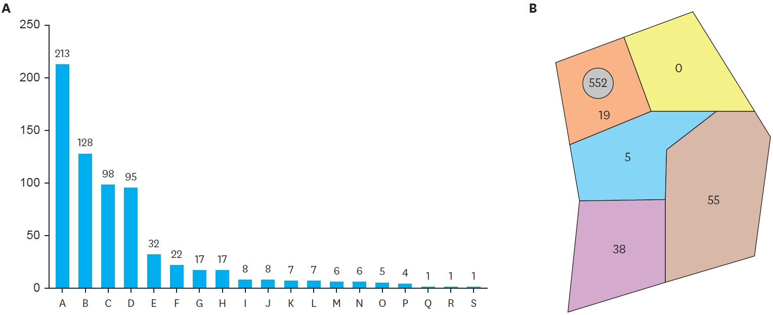
The Korean Association of Pediatric Surgeons (KAPS) performed a nationwide survey on sacrococcygeal teratoma in 2018.

Methods

The authors reviewed and analyzed the clinical data of patients who had been treated for sacrococcygeal teratoma by KAPS members from 2008 to 2017.

Results

A total of 189 patients from 18 institutes were registered for the study, which was the first national survey of this disease dealing with a large number of patients in Korea. The results were discussed at the 34th annual meeting of KAPS, which was held in Jeonju on June 21–22, 2018.

Conclusions

We believe that this study could be utilized as a guideline for the treatment of sacrococcygeal teratoma to diminish pediatric surgeons' difficulties in treating this disease and thus lead to better outcomes.

  • 10 View
  • 0 Download
[English]
Clinical Analysis of Foreign Bodies in Gastrointestinal Tract in Children
Eunsoo Choi, Hyo Gyun Lee, Soo Jin Na Choi, Sang Young Chung
J Korean Assoc Pediatr Surg 2014;20(1):12-16.   Published online June 30, 2014
DOI: https://doi.org/10.13029/jkaps.2014.20.1.12

Foreign body ingestion is a common problem among paediatric populations. Most of the ingested foreign bodies spontaneously pass through the gastrointestinal tract, but approximately less then 10% of them remain without being discharged, and trigger complications. Therefore, proper evaluation and treatment according to the situation is required. In this study, clinical progress and complications were analyzed according to the clinical features and treatment in children who ingested foreign bodies. Among pediatric patients under 18 who were admitted to Chonnam National University Hospital after ingesting foreign bodies between January 2008 to June 2012, only the patients who had their foreign body in the gastrointestinal tract were included in this study. Based on medical records, age, type of foreign body, time spent till admission, and whether the endoscopy was done or not, complication were researched retrospectively. According to symptoms and plain abdomen X-ray findings, treatment was chosen and conducted among endoscopy, observation and emergency operation. Among 273 patients, 9 (3.3%) of them had surgical removal. Seven (2.6%) of them had an emergency operation on the day of admission, and the rest 2 (0.7%) had operation during observation. Removal through initial endoscopic approach was tried in 157 (57.5%) patients. Eleven (70.8%) of them had their foreign body removed at the initial trial, and 5 (4.9%) of them at the second trial. Among 109, who were on observation status, 9 (8.3%) of them needed endoscopic removal, and 2 (1.8%) of them suffered from surgical removal. It is thought to be better to approach slowly considering the type, size and symptoms in foreign body ingestion of pediatric patients, rather than immediate and invasive removal.

  • 8 View
  • 0 Download
[English]
Multiple Tuberculous Splenic Abscesses in a Child
Chan Yong Park, Soo Jin Na Choi, Sang Young Chung, Shin Kon Kim
J Korean Assoc Pediatr Surg 2006;12(2):244-250.   Published online December 31, 2006
DOI: https://doi.org/10.13029/jkaps.2006.12.2.244

Splenic abscess is a rare clinical condition with a reported incidence of 0.14 % to 0.70 % in various autopsy series. Primary tuberculosis of the spleen as a cause of splenic abscess is even rarer, especially in the antitubercular era. Infants and children have a higher predisposition to extra-pulmonary tuberculosis than adults and tend to develop severe extra-pulmonary disease such as miliary tuberculosis and meningitis. The diagnosis of tuberculosis in infants and children can be difficult because of nonspecific symptoms and clinical findings. Computed tomography establishes the diagnosis of splenic abscess and demonstrates the number and location of abscesses. Splenectomy is the standard of care in most clinical setting. We present a 4-year-old girl who had multiple tuberculosis splenic abscesses and was treated successfully with splenectomy.

  • 4 View
  • 0 Download
[English]
Outcome of the Teratoma in Pediatric Surgical Patients
Ho Kyun Lee, Kyung Sub Park, Soo Jin Na Choi, Shin Kon Kim, Sang Young Chung
J Korean Assoc Pediatr Surg 2003;9(2):94-97.   Published online December 31, 2003
DOI: https://doi.org/10.13029/jkaps.2003.9.2.94

The teratoma is a unique complex neoplasm and is one of the most frequent pediatric tumors originated from the extragonadal germ cells. Mature teratoma is composed of mature differentiated tissues, while immature teratoma always contains embryonic tissues of variable degrees of immaturity, especially in the neuroepithelial elements. Diagnosis of teratoma is relatively easy by conventional radiologic study, but the immaturity can be identified only by histopathological examination. Between January 1993 to December 2002, 63 cases of teratoma were operated and analysed retrospectively at the Chonnam University Hospital Female to male ratio was about 3:1 and age distribution was relatively even. Among 63 cases, gonadal teratoma was the most common (52.4%), followed by sacrococcygeal (25.4%), retroperitoneal (9.5%) and mediastinal teratoma (9.5%). Fifty-six cases were mature teratomas and seven were immature teratomas. Alpha-fetoprotein (AFP) was elevated in 4 of 6 immature cases, but in 2 of 51 mature ones Elevated AFP progressively returned to normal range by 1 month after operation in all. Complete excision of the mass was performed, and major complication was not noticed. In five immature cases, PEB chemotherapy (Cisplatin, Etoposide, Bleomycin) was performed. Two of 2 cases in histological grade II were well tolerated to the aggressive chemotherapy. One of three cases in grade III expired due to severe bone marrow depression, and two of them expired by tumor recurrences. In conclusion, immature teratoma in histological grade III showed high potentiality of recurrence. Therefore, postoperative chemotherapy has to be applied to the high graded immature tumors.

  • 8 View
  • 0 Download
[English]
Gastroduodenal Intussusception due to Gastric Submucosal Hemangiomatosis
Soo Jin Na Choi, Sang Young Chung, Shin Kon Kim, Sang Woo Jung
J Korean Assoc Pediatr Surg 2000;6(2):149-152.   Published online December 31, 2000
DOI: https://doi.org/10.13029/jkaps.2000.6.2.149

Gastroduodenal intussusception, an invagination of a part of the gastric wall through the pyloric canal into the duodenum is a rare condition. Gastroduodenal intussusception is caused by a mobile usually benign gastric tumor. However, gastroduodenal intussusception by gastric submucosal hemangiomatosis is not documented. We have managed a case of gastric submucosal tumor leading to gastroduodenal intussusception in 2 years and 10 months old boy. The tumor was 10 × 5 × 3 cm in size in posterior wall of gastric antrum. Laparotomy, manual reduction of the intussusception, and wedge resection of posterior gastric wall including the tumor were performed. Pathologic diagnosis was a submucosal hemangiomatosis.

  • 7 View
  • 0 Download
[English]
A Clinical Review of Ten Years' Pediatric Surgical Experience
Soo Jin Na Choi, Sang Young Chung, Shin Kon Kim
J Korean Assoc Pediatr Surg 1998;4(2):110-116.   Published online December 31, 1998
DOI: https://doi.org/10.13029/jkaps.1998.4.2.110

This is a clinical review of 2,191 pediatric surgical patients under the age of 15 years, operated upon at the Division of Pediatric Surgery, Department of Surgery, Chonnam University Hospital from January 1988 to December 1997. The total number of operations in the pediatric age for all specialties were 13,144(13.2 %). The total operations including those performed on adults were 99,555. The most common age group operated upon was under 5 year of age(44.4 %). The number of operations in Division of General Pediatric Surgery were 2,191(16.7 %) out of total 13,144 operations in all pediatric specialties. The patients under 1 year of age in general pediatric surgery was 42.9 %(941/2,191). The most common diseases in neonates were anorectal malformation(20.6 %) and hypertrophic pyloric stenosis( 20.3 %). Infants older than neonates most commonly were operated upon for inguinal hernia(32.4 %) and intussusception(19.6 %). The total mortality rate in the neonatal intensive care unit was 31.3 %. Gastroschisis had the highest mortality.

  • 4 View
  • 0 Download

Case Report

[English]
A Case of Conjoined Twins: Omphalopagus
Soo Jin Na Choi, Sang Yong Chung, Shin Kon Kim
J Korean Assoc Pediatr Surg 1997;3(2):164-167.   Published online December 31, 1997
DOI: https://doi.org/10.13029/jkaps.1997.3.2.164

Conjoined twins are one of the rarest and most challenging congenital anomalies in pediatric surgery. Successful surgical separation is difficult because it the majority of conjoined twins in is technically typical to separate shared vital organs successfully. The timing of separation is variable, but separation is usually delayed until such infants are relatively mature(i.e, 9-12 months of age). Operative survival was 50 % in the neonatal period, but 90 % in those over 4 months of age. The present case was successfully separated early beacase of cardiac problems in one of the twins. These twins were omphalopagus and only the liver was shared through a bridge. The vascular and biliary trees were independent from each other. Successful surgical separation was on 11th day of life, but one died of sepsis 18 days ofter operation.

  • 6 View
  • 0 Download