ABSTRACT
Undifferentiated embryonal sarcoma of the liver (UESL) is a rare and highly aggressive malignant mesenchymal neoplasm that occurs mainly in school-aged children. Preoperative diagnosis is challenging because clinical symptoms are nonspecific and imaging findings can vary substantially. Despite these diagnostic difficulties, survival has improved markedly with modern multimodal treatment. A previously healthy 10-year-old girl presented with acute right upper quadrant abdominal pain. Laboratory findings were largely unremarkable, and the alpha-fetoprotein level was within the normal range. Contrast-enhanced abdominal computed tomography and magnetic resonance imaging showed a large heterogeneous cystic mass in the right hepatic lobe, with internal hemorrhage, hemorrhage-fluid levels, and enhancing solid components. Right hepatectomy was performed, and complete tumor resection was achieved. Histopathological evaluation revealed a high-grade malignant mesenchymal tumor with pathological and immunohistochemical features consistent with UESL. The patient subsequently received adjuvant chemotherapy. At 21 months after surgery, she remained disease-free, with no radiological or clinical evidence of recurrence. This case highlights the diagnostic value of characteristic imaging findings and the therapeutic importance of complete resection followed by adjuvant chemotherapy. Early recognition and coordinated multidisciplinary management are essential for improving prognosis in children with this rare hepatic malignancy.
-
Keywords: Undifferentiated embryonal sarcoma; Liver; Child; Case reports
INTRODUCTION
Undifferentiated embryonal sarcoma of the liver (UESL) is a rare malignant mesenchymal tumor that occurs mainly in school-aged children, particularly those 6 to 10 years of age [
1,
2]. It is the third most common malignant hepatic tumor in children, accounting for approximately 9% to 15% of pediatric malignant liver tumors after hepatoblastoma and hepatocellular carcinoma [
3]. UESL is a distinct clinicopathological entity characterized by aggressive biological behavior and historically poor outcomes. Because its clinical manifestations are nonspecific, affected patients often lack distinctive diagnostic features. Routine laboratory findings and serum tumor marker levels are also frequently unremarkable, which can delay diagnosis or lead to misdiagnosis as a benign pediatric liver tumor [
4]. Surgical resection alone was historically associated with poor prognosis, frequent recurrence, and early mortality. However, survival has improved substantially with multimodal treatment strategies that combine complete surgical resection with multiagent chemotherapy [
5,
6]. Here, we describe a case of UESL in a 10-year-old girl who presented with acute abdominal pain and was successfully treated with complete surgical resection followed by adjuvant chemotherapy. We also review the relevant literature to contextualize current diagnostic and therapeutic considerations for this rare pediatric hepatic malignancy.
CASE REPORT
Ethics statement
Written informed consent for publication of the case was obtained from the patient’s parents. This case report was prepared in accordance with the principles of the Declaration of Helsinki.
Patient information
A previously healthy 10-year-old girl presented to the emergency department of our institution with a 1-day history of right upper quadrant abdominal pain. She had no relevant personal or family medical history. On arrival, she reported severe right upper quadrant pain. Her vital signs were as follows: blood pressure, 132/91 mmHg; heart rate, 107 beats/min; respiratory rate, 18 breaths/min; and body temperature, 37.3 °C. Physical examination showed mild right upper quadrant tenderness, with no other remarkable findings.
Clinical and diagnostic findings
Initial laboratory testing showed a white blood cell count of 9,640/mm³, hemoglobin level of 12.9 g/dL, platelet count of 279,000/mm³, C-reactive protein level of 0.05 mg/dL, erythrocyte sedimentation rate of 3 mm/hr, alpha-fetoprotein (AFP) level of 4.9 ng/mL, and cancer antigen 125 level of 12.2 U/mL. Liver function testing showed aspartate aminotransferase and alanine aminotransferase levels of 43 and 50 U/L, respectively. The alkaline phosphatase level was 233 U/L, total bilirubin level was 0.54 mg/dL, total protein level was 6.8 g/dL, and albumin level was 4.3 g/dL.
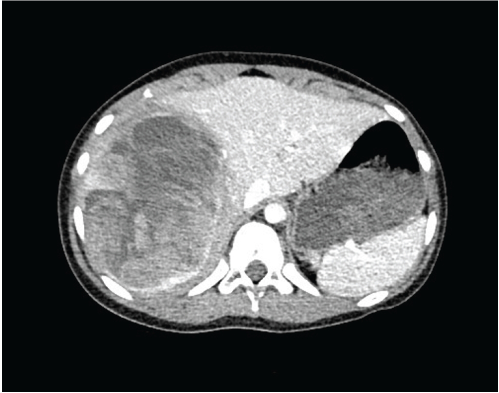
Contrast-enhanced abdominal computed tomography (CT) revealed a large heterogeneous mass measuring 13.0×11.6×9.2 cm and involving the right anterior and posterior hepatic segments. The lesion was predominantly water-attenuated and showed suspected acute hemorrhagic components and peripheral enhancement (
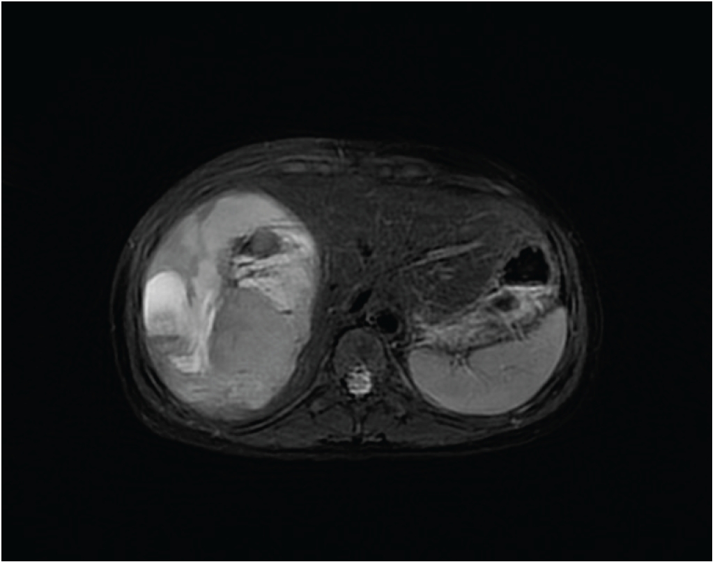
Fig. 1). Contrast-enhanced abdominal magnetic resonance imaging (MRI) demonstrated a large multiseptated cystic mass measuring 11.5×10.7×8.7 cm in the right hepatic lobe, with enhancing solid components and multiple hemorrhage-fluid levels. The lesion was hyperintense on diffusion-weighted imaging, and the overall findings were considered most consistent with UESL. The mass compressed and displaced the right hepatic vein, inferior vena cava, and right portal vein (
Fig. 2).
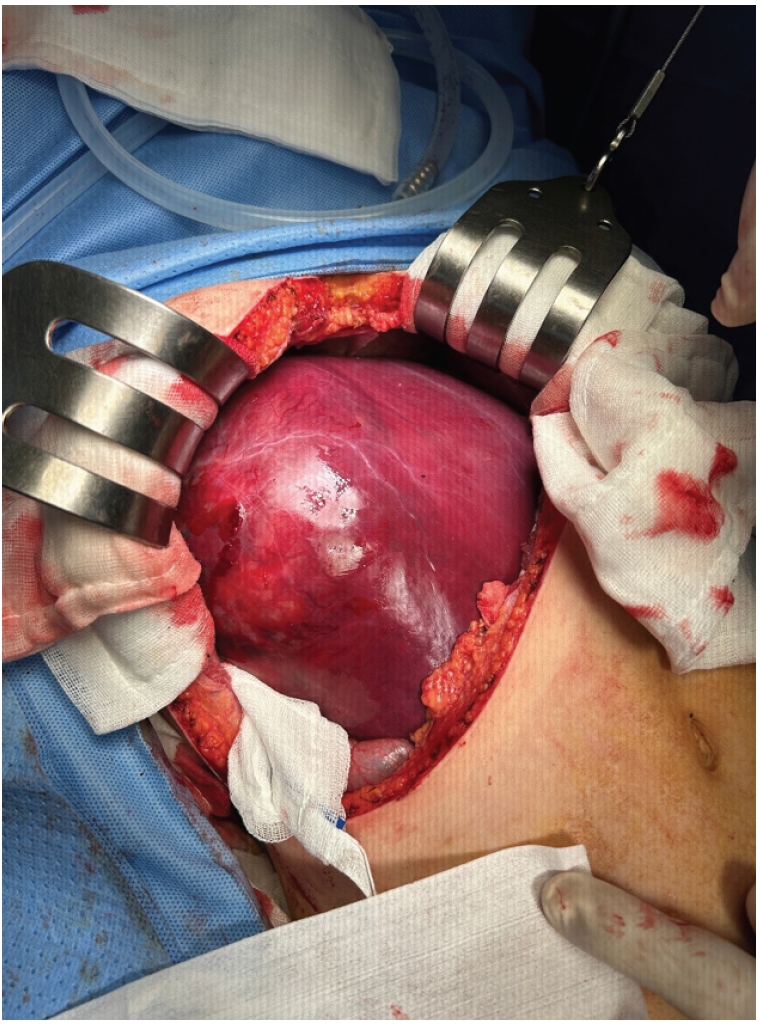
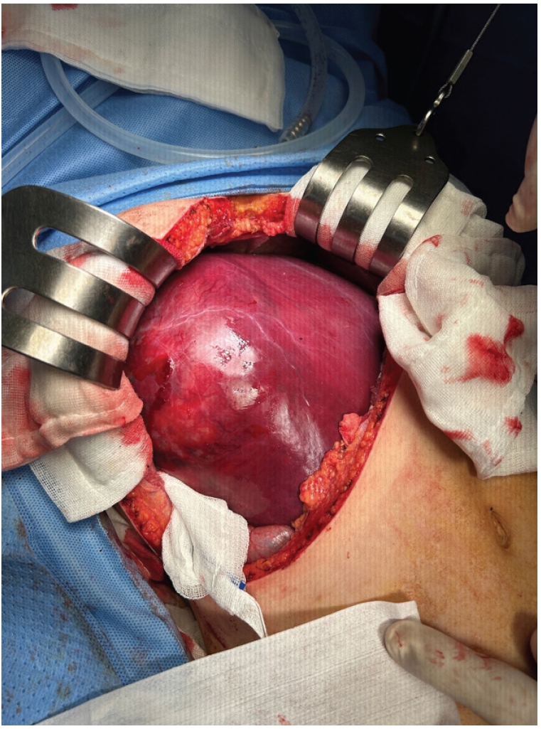
On the basis of these findings, surgical resection was planned. An extended right subcostal incision was made, and intraoperative exploration revealed a large mass occupying the right hepatic lobe (
Fig. 3). After the falciform ligament was divided, the right liver was fully mobilized. The portal triad was secured with umbilical tape, and the right portal vein and right hepatic artery were separately encircled and controlled with vessel loops. Intraoperative ultrasonography was used to define the tumor margins. After ischemic color change of the right hepatic lobe was confirmed, right hepatectomy was performed along the middle hepatic vein using a Cavitron ultrasonic surgical aspirator and an advanced bipolar energy device (Thunderbeat, Olympus).
Gross examination showed a right hepatectomy specimen measuring 17.5×14.0×9.5 cm and weighing 804 g. A single nodular tumor measuring 12.6×10.0×8.0 cm was present in the right hepatic lobe. The tumor showed no capsule formation or serosal infiltration. The surgical resection margin was negative, with a tumor-free distance of 1.5 cm. No gross vascular or bile duct invasion was identified. Microscopically, the tumor contained internal septa but lacked a capsule. There was no evidence of serosal infiltration, microvascular invasion, bile duct invasion, or satellite nodules. Necrosis involved approximately 10% of the tumor, and high mitotic activity with atypical mitotic figures was observed. Intracytoplasmic hyaline globules were present and were positive on periodic acid–Schiff staining. Immunohistochemical analysis showed positivity for vimentin, desmin, CD10, and glutamine synthetase, with focal positivity for hepatocyte antigen and glypican-3. The tumor was negative for β-catenin, cytokeratin 7, cytokeratin 19, c-kit, smooth muscle actin, AFP, S-100 protein, and CD34. INI-1 (BAF47) expression was preserved, without loss of nuclear staining. On the basis of the histopathological and immunohistochemical findings, the tumor was diagnosed as UESL.
Therapeutic intervention
Postoperatively, the patient was referred to the pediatric hematology-oncology department and received adjuvant chemotherapy according to the Children’s Oncology Group ARST0332 protocol (Arm C). The regimen comprised ifosfamide and doxorubicin, administered in seven cycles at 3- to 4-week intervals.
Follow-up and outcomes
The patient tolerated chemotherapy well, had no major complications, and was discharged in good condition. At the most recent follow-up, 21 months after surgery, she remained free of recurrence and continued outpatient surveillance.
DISCUSSION
UESL is a rare, high-grade malignant mesenchymal tumor that occurs mainly in school-aged children, especially those 6 to 10 years of age [
1,
2]. Since its landmark description by Stocker and Ishak in 1978, UESL has been regarded as the third most common malignant hepatic tumor in children, after hepatoblastoma and hepatocellular carcinoma, accounting for approximately 9% to 15% of pediatric malignant liver tumors [
2,
6]. Historically, UESL carried a poor prognosis, with a median survival of less than 1 year when treated with surgery alone. Over the past two decades, however, clinical outcomes have improved substantially, largely because of advances in multimodal treatment [
5,
6].
Because the clinical manifestations of UESL are nonspecific, affected patients may present with abdominal pain, fever, or an abdominal mass. Laboratory findings often provide limited diagnostic guidance, and AFP levels typically remain within the normal range. Consequently, early diagnosis and differentiation of UESL from other pediatric hepatic malignancies remain challenging. These features were reflected in the present case: the patient presented with acute right upper quadrant abdominal pain, while the serum AFP level was normal.
Imaging is central to preoperative suspicion for UESL. On ultrasonography, UESL typically appears as a hyperechoic, solid, heterogeneous tumor. CT helps characterize tumor location, boundaries, and relationships with adjacent vascular structures. On CT, UESL may appear as a solitary, well-defined, predominantly hypoattenuated mass; intratumoral serpiginous vessels have also been reported as an important diagnostic feature [
7]. MRI is particularly valuable for evaluating pediatric hepatic masses because it provides superior soft tissue contrast, permits the use of hepatobiliary contrast agents, and avoids ionizing radiation. MRI also helps assess vascular invasion, biliary obstruction, and hilar adenopathy. On MRI, UESL typically appears as a large, well-defined, multiseptated mass with hyperintensity on T2-weighted images and iso- to hypointensity on T1-weighted images [
6].
Preoperative diagnosis remains difficult because the clinical and radiological characteristics of UESL frequently overlap with those of other hepatic lesions. Mesenchymal hamartoma usually occurs in infants younger than 2 years and typically presents as a benign multiloculated cystic mass. Hepatoblastoma generally occurs in younger children and is more commonly associated with an elevated serum AFP level, calcification, and enhancing solid components. Complicated hepatic cysts or abscesses may also mimic UESL, but they are more likely to be accompanied by inflammatory symptoms, surrounding inflammatory changes, or a clinical history suggestive of infection or intracystic hemorrhage. Careful differential diagnosis is therefore essential, and ultrasound-guided biopsy may be important for establishing a definitive diagnosis. In addition, UESL is recognized as a highly friable tumor, and preoperative rupture has been reported in approximately 22% of cases [
8].
Microscopically, UESL is characterized by pleomorphic sarcomatous proliferation composed of poorly differentiated mesenchymal elements, with alternating loose and densely cellular areas surrounding bile ducts. Periodic acid–Schiff-positive cytoplasmic hyaline globules and mitotic activity, including abnormal mitotic figures, are commonly observed [
9].
Surgical resection remains the cornerstone of treatment for UESL. However, because of its malignant biology and rapid growth, UESL often presents as a large tumor that compresses adjacent organs and distorts major hepatic vascular structures, making resection technically demanding. In the present case, a definitive preoperative diagnosis could not be established on the basis of imaging alone. However, abdominal CT and MRI demonstrated a localized hepatic mass without radiological evidence of direct invasion into adjacent organs, major vascular involvement, or distant metastasis. Complete resection with an adequate surgical margin was therefore considered technically feasible. Because surgical resection could provide both definitive histological diagnosis and local tumor control, the patient underwent upfront resection without preoperative biopsy or neoadjuvant chemotherapy. Xiu et al. [
10] reported that three-dimensional reconstruction may help delineate the spatial relationship between the tumor and major blood vessels, while simulated surgery may support preoperative assessment of resectability. Nevertheless, surgery alone has historically been associated with poor prognosis, high recurrence rates, and early mortality. More recently, survival has improved significantly with multimodal treatment strategies combining complete surgical resection and multiagent chemotherapy [
11]. In a National Cancer Database analysis, Ziogas et al. [
12] reported a 5-year overall survival rate of 84.4% among pediatric patients with UESL. Several chemotherapeutic agents, including ifosfamide, etoposide, carboplatin, and doxorubicin, have been investigated for UESL treatment [
6]. In the present case, postoperative chemotherapy was administered according to the ARST0332 protocol, based on its established use in soft tissue sarcoma treatment. However, because no UESL-specific chemotherapy regimen has been established, its use in UESL remains an extrapolation from soft tissue sarcoma treatment strategies. Chemotherapy is generally administered in the neoadjuvant or adjuvant setting. In patients with a large tumor burden at diagnosis, neoadjuvant chemotherapy may reduce tumor volume and facilitate delayed surgical resection. It may also be considered when upfront complete resection is technically difficult because of tumor proximity to major hepatic vessels [
6]. When complete resection is not feasible because of extensive vascular involvement or insufficient future liver remnant, liver transplantation has emerged as a potential curative option. Rolfes et al. [
13] reported favorable outcomes after liver transplantation in patients with UESL, with survival comparable to that of patients undergoing transplantation for hepatoblastoma or nonmalignant indications. On the basis of these findings, they suggested that liver transplantation should be considered for patients with unresectable, localized UESL.
In conclusion, this case highlights the importance of early recognition of characteristic imaging findings, definitive surgical management aimed at complete resection, and adjuvant chemotherapy for achieving favorable outcomes in pediatric UESL. At 21 months after surgery, the patient remained disease-free, consistent with the favorable short- to mid-term outcomes described in recent literature. Although recurrence is uncommon after complete resection and multimodal therapy, continued long-term surveillance remains essential.
NOTES
-
Authors’ contributions
Conceptualization: all authors. Formal analysis: all authors. Writing–original draft: all authors. Writing–review & editing: all authors. All authors read and approved the final manuscript.
-
Conflict of interest
None.
-
Funding
None.
-
Data availability
Not applicable.
Fig. 1.Contrast-enhanced abdominal computed tomography showing a large heterogeneous mass measuring 13.0×11.6×9.2 cm and involving the right anterior and posterior hepatic segments. The lesion is predominantly water-attenuated, with suspected areas of acute hemorrhage and peripheral enhancement.
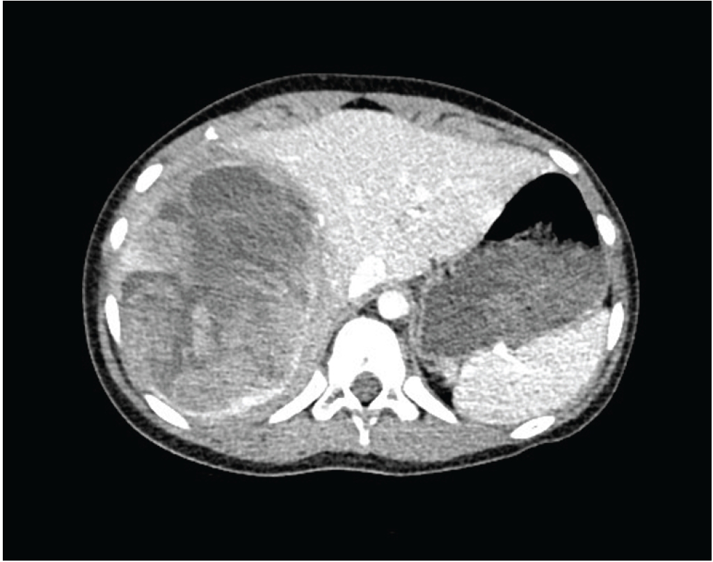
Fig. 2.Contrast-enhanced abdominal magnetic resonance imaging demonstrating a large multiseptated cystic mass measuring 11.5×10.7×8.7 cm in the right hepatic lobe, with enhancing solid components and multiple hemorrhage-fluid levels.
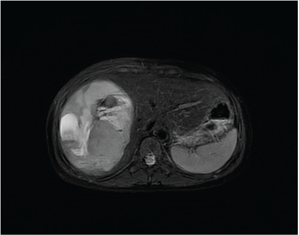
Fig. 3.Intraoperative image showing a large mass occupying the right hepatic lobe.
REFERENCES
- 1. Halefoglu AM, Oz A. Primary undifferentiated embryonal sarcoma of the liver misdiagnosed as hydatid cyst in a child: a case report and review of the literature. JBR-BTR 2014;97:248-50.
- 2. van Noesel MM. Rare pediatric tumors benefit from large clinical trials: the story of undifferentiated embryonal sarcoma of the liver. Cancer 2024;130:2582-5.
- 3. Shi Y, Rojas Y, Zhang W, et al. Characteristics and outcomes in children with undifferentiated embryonal sarcoma of the liver: a report from the National Cancer Database. Pediatr Blood Cancer 2017;64:e26272.
- 4. Zhang C, Jia CJ, Xu C, Sheng QJ, Dou XG, Ding Y. Undifferentiated embryonal sarcoma of the liver: clinical characteristics and outcomes. World J Clin Cases 2020;8:4763-72.
- 5. Li J, He L, Wu Q, Zhou J, Zhou L, Li T. Undifferentiated embryonal sarcoma of the liver in pediatric patients: a single-institution retrospective case series. Transl Pediatr 2024;13:784-90.
- 6. Lin WY, Wu KH, Chen CY, et al. Treatment of undifferentiated embryonal sarcoma of the liver in children. Cancers (Basel) 2024;16:897.
- 7. Gabor F, Franchi-Abella S, Merli L, Adamsbaum C, Pariente D. Imaging features of undifferentiated embryonal sarcoma of the liver: a series of 15 children. Pediatr Radiol 2016;46:1694-704.
- 8. Kastenberg ZJ, Short SS, Riehle KJ, et al. Management of undifferentiated embryonal sarcoma of the liver: a Pediatric Surgical Oncology Research Collaborative study. Pediatr Blood Cancer 2024;71:e30975.
- 9. Mohin M, Mukherjee R, Dewan L, Karan S, Chatterjee U. Cytological findings in a case of undifferentiated embryonal sarcoma of liver in a child: report of a case with review of literature. Cytopathology 2024;35:416-20.
- 10. Xiu W, Li T, Liu J, et al. Undifferentiated embryonal sarcoma of the liver in children: our experience in four difficult cases and three-dimensional practical exploration. World J Surg Oncol 2024;22:222.
- 11. Gao J, Fei L, Li S, et al. Undifferentiated embryonal sarcoma of the liver in a child: a case report and review of the literature. Oncol Lett 2013;5:739-42.
- 12. Ziogas IA, Zamora IJ, Lovvorn HN III, Bailey CE, Alexopoulos SP. Undifferentiated embryonal sarcoma of the liver in children versus adults: a National Cancer Database analysis. Cancers (Basel) 2021;13:2918.
- 13. Rolfes PS, Yoeli D, Feldman AG, Adams MA, Wachs ME, Boster JM. Modern-era successful liver transplantation outcomes in children with hepatic undifferentiated embryonal sarcoma. Pediatr Transplant 2024;28:e14645.