ABSTRACT
Rectal polyps are relatively common in children and most often present as painless rectal bleeding after defecation. By contrast, rectal prolapse caused by an underlying rectal polyp is uncommon, especially in pediatric patients. Because intermittent prolapse may have resolved by the time of clinical examination, the diagnosis can be difficult to establish. A previously healthy 1-year-old boy was referred to the pediatric surgery outpatient clinic because of a 6-month history of intermittent protrusion of a mass through the anus after defecation. The episodes initially occurred approximately three times per day but gradually increased to six times per day. Physical examination showed no definite perianal abnormality; however, a photograph provided by the patient’s mother showed a protruding anorectal mass, raising suspicion for rectal prolapse associated with a polyp. Colonoscopy revealed a large pedunculated rectal polyp, measuring approximately 47×34×25 mm, with a short stalk located approximately 15 cm proximal to the anal verge. The patient underwent transanal excision under general anesthesia. Histopathological examination confirmed a Peutz-Jeghers-type hamartomatous polyp. The postoperative course was uneventful, and no recurrence was observed during 3 years of follow-up. Rectal polyps should be considered in the differential diagnosis of recurrent rectal prolapse in infants and young children. Caregiver-provided photographs can help establish the diagnosis when the lesion is not visible during examination. In selected pediatric patients with large rectal hamartomatous polyps presenting with recurrent prolapse, transanal excision may provide safe and effective treatment.
-
Keywords: Rectal prolapse; Polyps; Child; Case reports
INTRODUCTION
Rectal polyps in children commonly cause lower gastrointestinal symptoms, particularly painless rectal bleeding after defecation [
1]. Other possible manifestations include mucus discharge, anemia, and, rarely, protrusion through the anus. Colonoscopy is usually used to establish the diagnosis because it permits direct visualization, assessment of the number and location of polyps, and therapeutic removal. Histologically, most pediatric colorectal polyps are juvenile polyps; however, other hamartomatous lesions, including Peutz-Jeghers-type polyps, may also occur.
Rectal prolapse is also common in infants and young children and is generally attributed to the relatively vertical orientation of the rectum, increased sigmoid colon mobility, and immature pelvic floor support [
2]. Most cases resolve with conservative management [
3]. However, recurrent rectal prolapse accompanied by a protruding mass is unusual and should prompt evaluation for an underlying rectal lesion.
Rectal prolapse caused by a rectal polyp is rare, particularly in children. Although some adult cases have been reported [
4-
6], pediatric cases remain uncommon [
7-
9]. This report describes a 1-year-old boy with recurrent rectal prolapse caused by a large Peutz-Jeghers-type rectal hamartomatous polyp that was successfully treated by transanal excision, and it briefly reviews the relevant literature.
CASE REPORT
Ethics statement
Written informed consent for publication of the case was obtained from the patient’s parents. This case report was prepared in accordance with the principles of the Declaration of Helsinki.
Patient information
A previously healthy 1-year-old boy was referred to the pediatric surgery outpatient clinic because of a 6-month history of intermittent protrusion of a mass through the anus after defecation. The prolapse initially occurred approximately three times daily and gradually increased to six episodes per day. He had no relevant personal or family medical history.
Clinical and diagnostic findings
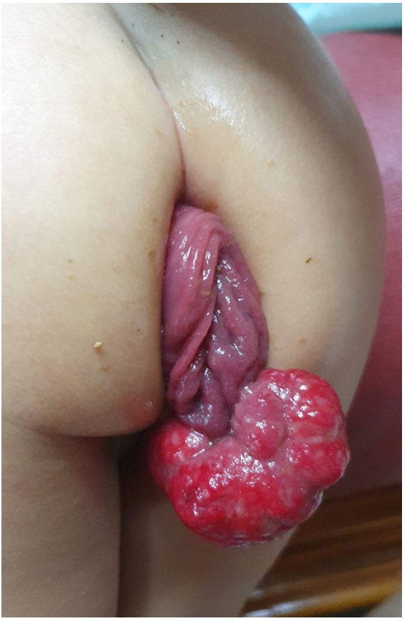
Outpatient physical examination showed no definite abnormality. However, a photograph provided by the patient’s mother showed a protruding anorectal mass, raising suspicion for rectal prolapse associated with a polyp (
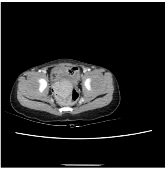
Fig. 1). On arrival, his vital signs were as follows: blood pressure, 104/62 mmHg; heart rate, 128 beats/min; respiratory rate, 24 breaths/min; and body temperature, 36.5 °C. Laboratory findings showed no significant abnormalities. Abdominal computed tomography (CT), performed as part of the evaluation for rectal prolapse, showed an approximately 4-cm polypoid mass in the upper rectum (
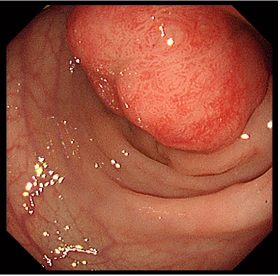
Fig. 2). The outer margin of the rectum was smooth, and there were no significantly enlarged regional lymph nodes, ascites, or other abdominal abnormalities. Colonoscopy revealed a large pedunculated rectal polyp measuring approximately 47×34×25 mm, with a short stalk located approximately 15 cm proximal to the anal verge (
Fig. 3). Careful examination of the remaining colorectal mucosa showed no additional synchronous polyps or other mucosal abnormalities.
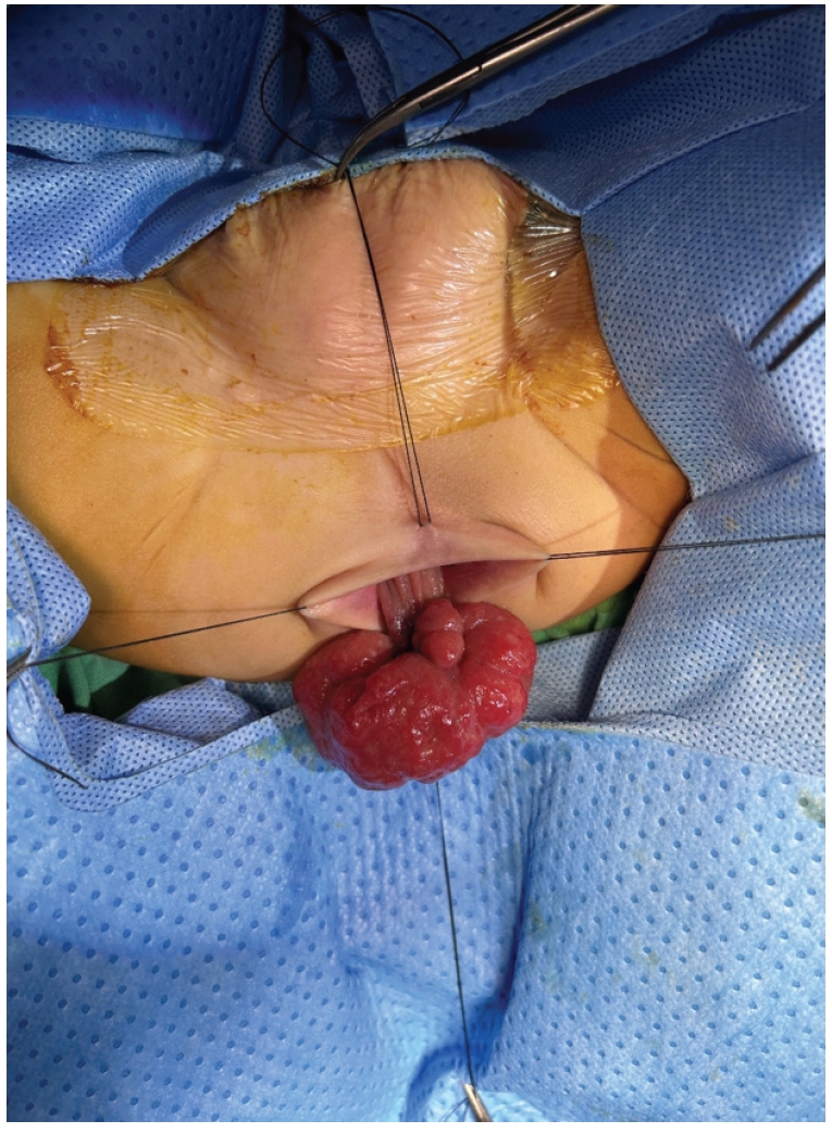
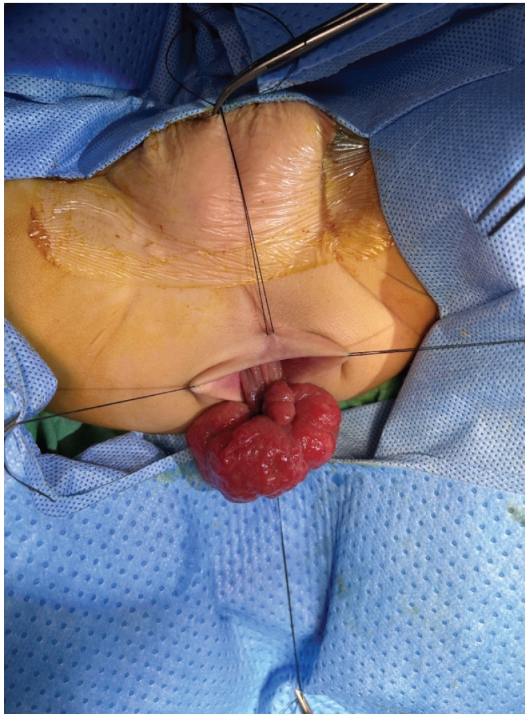
Because the polyp was large, conventional endoscopic polypectomy was considered technically difficult and potentially associated with bleeding or incomplete resection. Surgical resection was therefore performed because the polyp was associated with frequent rectal prolapse. Under general anesthesia, the patient was placed in the lithotomy position. Digital rectal examination identified a rectal polyp at the 11 o’clock position, approximately 10 cm above the dentate line. Manual pressure applied to the lower abdomen enabled downward traction of the rectum and transanal exposure of the polyp outside the anus (
Fig. 4). The polyp was excised transanally, and the mucosa was repaired with 3-0 black silk sutures. Histopathological examination confirmed a Peutz-Jeghers-type hamartomatous polyp.
The postoperative course was uneventful, with no complications. Further evaluation revealed no mucocutaneous pigmentation or additional gastrointestinal polyps, and no relevant family history was reported.
DISCUSSION
Rectal prolapse is relatively common in infants and young children and is usually associated with functional or anatomical factors such as constipation, diarrhea, malnutrition, increased straining during defecation, and pelvic floor immaturity [
2]. In most pediatric patients, conservative treatment that corrects predisposing factors and improves bowel habits is sufficient [
3]. However, frequent or recurrent prolapse, especially when accompanied by a protruding mass, should prompt evaluation for an underlying colorectal lesion.
In the present case, recurrent protrusion of a mass through the anus after defecation clinically mimicked rectal prolapse. Although outpatient physical examination showed no definite abnormality, the caregiver-provided photograph suggested rectal prolapse associated with a polyp. Subsequent colonoscopy and abdominal CT identified a large polypoid mass in the upper rectum. This case illustrates why caregiver-provided photographs should be reviewed when symptoms are intermittent and absent during examination.
Histopathological examination revealed a Peutz-Jeghers-type hamartomatous polyp. Peutz-Jeghers polyps are characterized by an arborizing smooth muscle core extending from the muscularis mucosae and covered by normal-appearing gastrointestinal epithelium. These polyps may occur as part of Peutz-Jeghers syndrome, which is associated with mucocutaneous pigmentation, a family history, multiple gastrointestinal hamartomatous polyps, and germline mutations in
STK11, also known as
LKB1 (liver kinase B1) [
6]. However, solitary Peutz-Jeghers-type polyps can also occur in patients without clinical features of the syndrome. Patients diagnosed with a Peutz-Jeghers-type hamartomatous polyp should therefore be assessed carefully for mucocutaneous pigmentation, relevant family history, and additional gastrointestinal polyps. Because
STK11 genetic testing was not performed after surgery in this patient, Peutz-Jeghers syndrome could not be completely excluded on a genetic basis.
Peutz-Jeghers-type polyps may develop anywhere in the gastrointestinal tract, including the colon and rectum. Symptoms often begin during childhood and may include abdominal pain, bleeding, anemia, obstruction, or intussusception. Prolapse of a polyp through the anus is uncommon but may occur in young children, in whom it can mimic rectal prolapse [
7-
9]. In infants and toddlers, immature pelvic support and increased straining during defecation may contribute to polyp protrusion.
Management of a prolapsing rectal polyp depends on the size and location of the polyp, the patient’s symptoms, and whether endoscopic or surgical removal is technically feasible. Endoscopic polypectomy is often considered when it can be performed safely and completely. In the present case, the polyp was large, located in the upper rectum, and associated with frequent prolapse. Manual pressure applied to the lower abdomen lowered the rectum enough to permit transanal delivery of the polyp, allowing excision and mucosal repair through a transanal approach. This approach enabled complete removal without abdominal surgery.
The postoperative course was uneventful, and no recurrence was observed during 3 years of follow-up. Although this is a single case, the outcome suggests that transanal excision may be an effective, minimally invasive option for selected pediatric patients with large rectal hamartomatous polyps presenting with recurrent prolapse.
In conclusion, rectal polyps should be included in the differential diagnosis of recurrent rectal prolapse or intermittent anal protrusion in infants and young children. Caregiver-provided photographs can be particularly useful when the lesion is not visible during examination. For large rectal Peutz-Jeghers-type hamartomatous polyps presenting with prolapse, transanal excision may be a safe and effective treatment in carefully selected cases.
NOTES
-
Authors’ contributions
All the work was done by JP.
-
Conflict of interest
None.
-
Funding
None.
-
Data availability
Not applicable.
Fig. 1.Caregiver-provided photograph showing a protruding anorectal mass suggestive of rectal prolapse associated with a polyp.
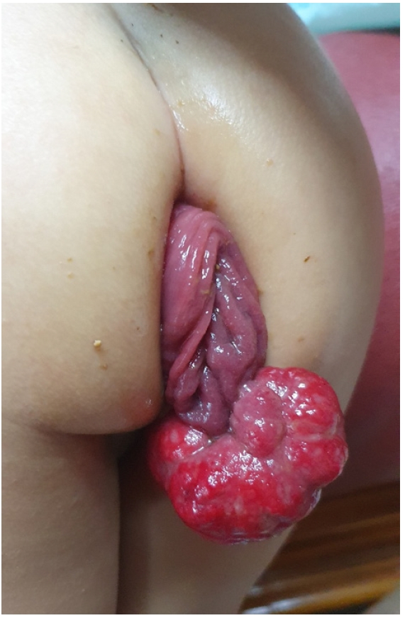
Fig. 2.Abdominal computed tomography image showing an approximately 4-cm polypoid mass in the upper rectum.
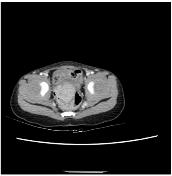
Fig. 3.Colonoscopic image showing a large, round, hyperemic rectal mass measuring approximately 5 cm and located 15 cm from the anal verge.
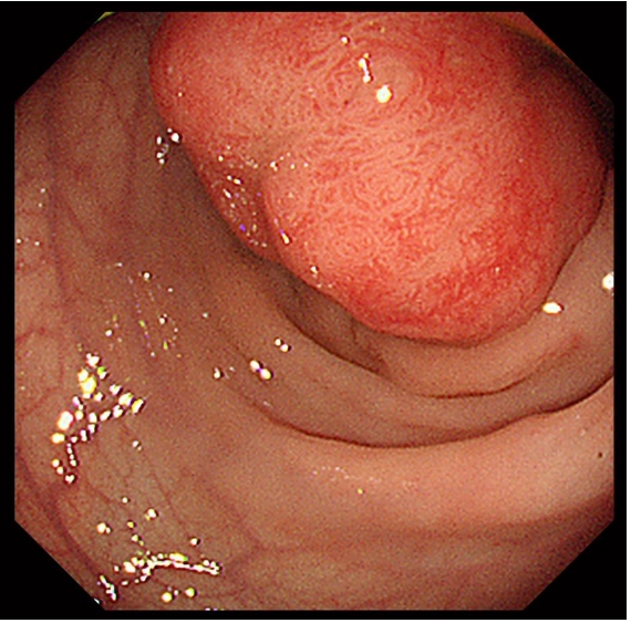
Fig. 4.Transanal exposure of the rectal polyp using 3-0 black silk traction sutures before excision.
REFERENCES
- 1. Wiseman J, Emil S. Minimal access surgical management of large juvenile polyps in children. J Pediatr Surg 2009;44:e9-13.
- 2. Short SS, Wynne EK, Zobell S, Gaddis K, Rollins MD. Most children experience resolution of idiopathic pediatric rectal prolapse with bowel management alone. J Pediatr Surg 2022;57:354-8.
- 3. Kodia K, Huerta CT, Perez EA. Rectal prolapse in the pediatric population-a narrative review of medical and surgical management. Transl Gastroenterol Hepatol 2024;9:47.
- 4. Khan AA, Cervera LF, Shihadeh S, Glotzer D. Large anal polyp disguised as rectal prolapse. Cureus 2024;16:e65193.
- 5. De Leon Murillo AD, Plaza Ricardo PA, Perez Garcia TP, Castro Rodriguez JM, Anaya Meza SF, Pezzano Molina E. The Altemeier procedure in the management of rectal prolapse secondary to a prolapsed rectal polyp: a valid alternative. Cureus 2025;17:e96216.
- 6. Cano-Contreras AD, Meixueiro-Daza A, Grube-Pagola P, Remes-Troche JM. Giant rectal polyp prolapse in an adult patient with the Peutz-Jeghers syndrome. BMJ Case Rep 2016;2016:bcr2016215629.
- 7. Arredondo Montero J, Carracedo Vega E, Razquin Lizarraga S, et al. Pediatric rectosigmoid atypical juvenile polyps presenting with rectal prolapse and acute bleeding: a case report and a comprehensive literature review. Pediatr Dev Pathol 2024;27:340-7.
- 8. Nabar S, Bittar K. Rectal polyp masquerading as rectal prolapse. J Pediatr Gastroenterol Nutr 2023;77:e68.
- 9. Kakiuchi T, Yoshiura M. Juvenile polyp presenting as prolapsed mass per rectum in a 5-year-old child. Clin Case Rep 2023;11:e7145.
Citations
Citations to this article as recorded by