Congenital mesoblastic nephroma (CMN) is the most common renal tumor of early infancy, and infants with renal masses suspicious for CMN often undergo upfront radical nephrectomy with proximal ureterectomy. Infective mimics, including renal abscess, acute lobar nephronia, and xanthogranulomatous pyelonephritis, are considerably less common, particularly when typical clinical signs of infection are absent or muted. We report the case of a 2-month-old boy with a painless, progressively enlarging left abdominal mass first noted at 20 days of age, accompanied by intermittent low-grade fever and constipation. Contrast-enhanced computed tomography showed an ill-defined, heterogeneously enhancing left renal mass with perinephric extension and regional lymphadenopathy, raising suspicion for CMN. The infant underwent radical left nephroureterectomy with splenic flexure resection, the latter of which was required because of dense inflammatory adhesions. Histopathological examination revealed a chronic suppurative renal abscess with palisading histiocytes and multinucleated giant cells, with no evidence of malignancy, fungal organisms, or mycobacteria. Postoperative recovery was uneventful, and the child remains well during follow-up. This case illustrates a rare but clinically important diagnostic pitfall: infective renal masses can masquerade as malignant renal tumors in early infancy, particularly when systemic inflammatory features are subtle.
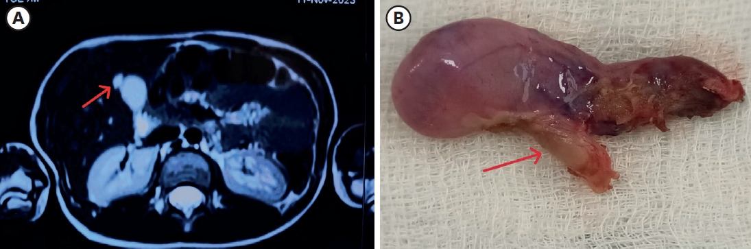
A ciliated foregut cyst is a rare developmental anomaly. It develops from the primitive foregut. It is usually located supra-diaphragmatically. Its localization in the gallbladder is very infrequent and has been sparsely reported. We report a rare case of a ciliated cyst of the gallbladder in an 11-year-old female, who presented with complaints of upper abdominal pain for 2 months. She was suspected to have gallbladder duplication or gallbladder diverticulum on imaging. The histopathology reported this anomaly as a ciliated foregut cyst. The ciliated cyst of the gallbladder is a benign congenital lesion. Abdominal ultrasonogram and computed tomography/magnetic resonance imaging are suggestive of a cystic lesion of the gallbladder. The definitive diagnosis is by histopathological examination. This is a rare clinicopathological condition in the pediatric age group. The recommended treatment is laparoscopic cholecystectomy. The role of conservative management has not been established due to the rarity of the condition.
Citations
Citations to this article as recorded by
Fundic Gallbladder Diverticulum Lesion in a Child Suspected to Be Congenital: A Case Report and Scoping Review Samentha Menager, Piero Farruggia, Salvatore Calderaro, Domenico Bivona, Giovanni Francesco Saia, Marco Guida, Antonino Levita, Chiara Marino, Federica Mescolo, Tiziana Guida Rutilio, Michelangelo Giovanni Sciortino, Barbara Torrente, Flavia Volpe, Giovan Pediatric Reports.2026; 18(5): 117. CrossRef



 First
 Prev